Ubiquitin mouse Monoclonal Antibody(5F1)
- Catalog No.:YM3636
- Applications:WB;IF;IHC
- Reactivity:Human;Rat;Mouse;Pig
- Target:
- UB
- Protein Name:
- Ubiquitin
- Human Gene Id:
- 7314
- Human Swiss Prot No:
- P62979;P62987;P62988
- Immunogen:
- Synthetic Peptide of Ubiquitin
- Specificity:
- Ubiquitin protein detects endogenous levels of Ubiquitin
- Formulation:
- Liquid in PBS containing 50% glycerol, 0.5% BSA and 0.02% sodium azide.
- Source:
- Monoclonal, Mouse
- Dilution:
- WB 1:1000-2000, IHC 1:100-200 IF 1:200
- Purification:
- The antibody was affinity-purified from mouse ascites by affinity-chromatography using specific immunogen.
- Concentration:
- 1 mg/ml
- Storage Stability:
- -15°C to -25°C/1 year(Do not lower than -25°C)
- Background:
- UBB (ubiquitin B) encodes ubiquitin, one of the most conserved proteins known. Ubiquitin has a major role in targeting cellular proteins for degradation by the 26S proteosome. It is also involved in the maintenance of chromatin structure, the regulation of gene expression, and the stress response. Ubiquitin is synthesized as a precursor protein consisting of either polyubiquitin chains or a single ubiquitin moiety fused to an unrelated protein. UBB consists of three direct repeats of the ubiquitin coding sequence with no spacer sequence. Consequently, the protein is expressed as a polyubiquitin precursor with a final amino acid after the last repeat. An aberrant form of this protein has been detected in patients with Alzheimer's disease and Down syndrome. Pseudogenes of UBB are located on chromosomes 1, 2, 13, and 17. Alternative splicing results in multiple transcript variants.
GRP94 Inhabits the Immortalized Porcine Hepatic Stellate Cells Apoptosis under Endoplasmic Reticulum Stress through Modulating the Expression of IGF-1 and Ubiquitin
Reprogramming Tumor-Associated Macrophages via ROS-Mediated Novel Mechanism of Ultra-Small Cu2−xSe Nanoparticles to Enhance Anti-Tumor Immunity. ADVANCED FUNCTIONAL MATERIALS 2021 Nov 12 IF Mouse Macrophages
LRRC19 expressed in the kidney induces TRAF2/6-mediated signals to prevent infection by uropathogenic bacteria. Nature Communications Nat Commun. 2014 Jul;5(1):1-12 WB Human HEK293T cell
High‐intensity interval training changes the expression of muscle RING‐finger protein‐1 and muscle atrophy F‐box proteins and proteins involved in the mechanistic target of rapamycin pathway and autophagy in rat skeletal muscle. EXPERIMENTAL PHYSIOLOGY 2019 Aug 29 WB Rat 1:1000 soleus muscle
Cui, Xinwen, et al. "HIIT changes the expression of MuRF1 and MAFBx proteins and proteins involved in the mTOR pathway and autophagy in rat skeletal muscle." Experimental Physiology.
- June 19-2018
- WESTERN IMMUNOBLOTTING PROTOCOL
- June 19-2018
- IMMUNOHISTOCHEMISTRY-PARAFFIN PROTOCOL
- June 19-2018
- IMMUNOFLUORESCENCE PROTOCOL
- September 08-2020
- FLOW-CYTOMEYRT-PROTOCOL
- May 20-2022
- Cell-Based ELISA│解您多样本WB检测之困扰
- July 13-2018
- CELL-BASED-ELISA-PROTOCOL-FOR-ACETYL-PROTEIN
- July 13-2018
- CELL-BASED-ELISA-PROTOCOL-FOR-PHOSPHO-PROTEIN
- July 13-2018
- Antibody-FAQs
- Products Images

- Immunohistochemical analysis of paraffin-embedded Human-Tonsil tissue. 1,Ubiquitin Mouse Monoclonal Antibody(5F1) was diluted at 1:200(4°C,overnight). 2, Sodium citrate pH 6.0 was used for antibody retrieval(>98°C,20min). 3,Secondary antibody was diluted at 1:200(room tempeRature, 30min). Negative control was used by secondary antibody only.
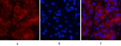
- Immunofluorescence analysis of Human-stomach-cancer tissue. 1,Ubiquitin Mouse Monoclonal Antibody(5F1)(red) was diluted at 1:200(4°C,overnight). 2, Cy3 labled Secondary antibody was diluted at 1:300(room temperature, 50min).3, Picture B: DAPI(blue) 10min. Picture A:Target. Picture B: DAPI. Picture C: merge of A+B
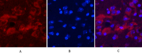
- Immunofluorescence analysis of Mouse-brain tissue. 1,Ubiquitin Mouse Monoclonal Antibody(5F1)(red) was diluted at 1:200(4°C,overnight). 2, Cy3 labled Secondary antibody was diluted at 1:300(room temperature, 50min).3, Picture B: DAPI(blue) 10min. Picture A:Target. Picture B: DAPI. Picture C: merge of A+B
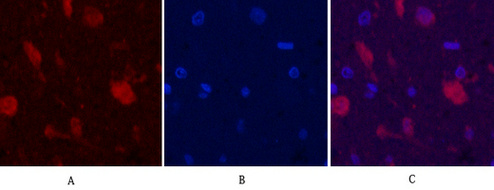
- Immunofluorescence analysis of Rat-brain tissue. 1,Ubiquitin Mouse Monoclonal Antibody(5F1)(red) was diluted at 1:200(4°C,overnight). 2, Cy3 labled Secondary antibody was diluted at 1:300(room temperature, 50min).3, Picture B: DAPI(blue) 10min. Picture A:Target. Picture B: DAPI. Picture C: merge of A+B
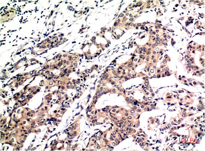
- Immunohistochemical analysis of paraffin-embedded Human Breast Carcinoma Tissue using Ubiquitin Mouse mAb diluted at 1:200.

- Western blot analysis of 1) Hela Cell Lysate, 2) 3T3 Cell Lysate, 3) Rat Brain Tissue Lysate using Ubiquitin Mouse mAb diluted at 1:1000.


