T-cadherin Polyclonal Antibody
- Catalog No.:YT4572
- Applications:WB;IHC;IF;ELISA
- Reactivity:Human;Mouse;Rat
- Target:
- T-cadherin
- Gene Name:
- CDH13
- Protein Name:
- Cadherin-13
- Human Gene Id:
- 1012
- Human Swiss Prot No:
- P55290
- Mouse Gene Id:
- 12554
- Mouse Swiss Prot No:
- Q9WTR5
- Immunogen:
- The antiserum was produced against synthesized peptide derived from human CDH13. AA range:331-380
- Specificity:
- T-cadherin Polyclonal Antibody detects endogenous levels of T-cadherin protein.
- Formulation:
- Liquid in PBS containing 50% glycerol, 0.5% BSA and 0.02% sodium azide.
- Source:
- Polyclonal, Rabbit,IgG
- Dilution:
- WB 1:500 - 1:2000. IHC 1:100 - 1:300. IF 1:200 - 1:1000. ELISA: 1:20000. Not yet tested in other applications.
- Purification:
- The antibody was affinity-purified from rabbit antiserum by affinity-chromatography using epitope-specific immunogen.
- Concentration:
- 1 mg/ml
- Storage Stability:
- -15°C to -25°C/1 year(Do not lower than -25°C)
- Other Name:
- CDH13;CDHH;Cadherin-13;Heart cadherin;H-cadherin;P105;Truncated cadherin;T-cad;T-cadherin
- Observed Band(KD):
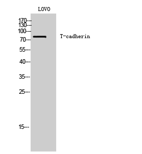
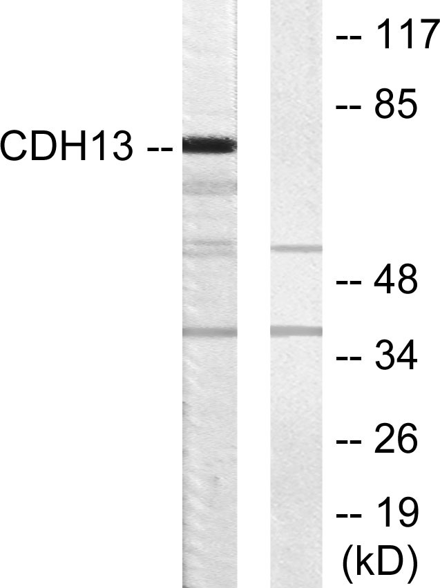
- 78kD
- Background:
- This gene encodes a member of the cadherin superfamily. The encoded protein is localized to the surface of the cell membrane and is anchored by a GPI moiety, rather than by a transmembrane domain. The protein lacks the cytoplasmic domain characteristic of other cadherins, and so is not thought to be a cell-cell adhesion glycoprotein. This protein acts as a negative regulator of axon growth during neural differentiation. It also protects vascular endothelial cells from apoptosis due to oxidative stress, and is associated with resistance to atherosclerosis. The gene is hypermethylated in many types of cancer. Alternative splicing results in multiple transcript variants encoding different isoforms. [provided by RefSeq, May 2011],
- Function:
- developmental stage:Expressed at higher levels in adult brain than in developing brain.,function:Cadherins are calcium dependent cell adhesion proteins. They preferentially interact with themselves in a homophilic manner in connecting cells; cadherins may thus contribute to the sorting of heterogeneous cell types. May act as a negative regulator of neural cell growth.,similarity:Contains 5 cadherin domains.,tissue specificity:Highly expressed in heart. In the CNS, expressed in cerebral cortex, medulla, hippocampus, amygdala, thalamus and substantia nigra. No expression detected in cerebellum or spinal cord.,
- Subcellular Location:
- Cell membrane; Lipid-anchor, GPI-anchor.
- Expression:
- Highly expressed in heart. In the CNS, expressed in cerebral cortex, medulla, hippocampus, amygdala, thalamus and substantia nigra. No expression detected in cerebellum or spinal cord.
- June 19-2018
- WESTERN IMMUNOBLOTTING PROTOCOL
- June 19-2018
- IMMUNOHISTOCHEMISTRY-PARAFFIN PROTOCOL
- June 19-2018
- IMMUNOFLUORESCENCE PROTOCOL
- September 08-2020
- FLOW-CYTOMEYRT-PROTOCOL
- May 20-2022
- Cell-Based ELISA│解您多样本WB检测之困扰
- July 13-2018
- CELL-BASED-ELISA-PROTOCOL-FOR-ACETYL-PROTEIN
- July 13-2018
- CELL-BASED-ELISA-PROTOCOL-FOR-PHOSPHO-PROTEIN
- July 13-2018
- Antibody-FAQs
- Products Images

- Western Blot analysis of LOVO cells using T-cadherin Polyclonal Antibody

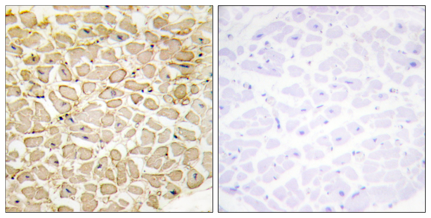
- Immunohistochemistry analysis of paraffin-embedded human heart tissue, using CDH13 Antibody. The picture on the right is blocked with the synthesized peptide.

- Western blot analysis of lysates from LOVO cells, using CDH13 Antibody. The lane on the right is blocked with the synthesized peptide.


