Versican Polyclonal Antibody
- Catalog No.:YT4874
- Applications:WB;IHC;IF;ELISA
- Reactivity:Human;Rat;Mouse;
- Target:
- Versican
- Fields:
- >>Cell adhesion molecules
- Gene Name:
- VCAN
- Protein Name:
- Versican core protein
- Human Gene Id:
- 1462
- Human Swiss Prot No:
- P13611
- Mouse Swiss Prot No:
- Q62059
- Immunogen:
- The antiserum was produced against synthesized peptide derived from human VCAN. AA range:532-581
- Specificity:
- Versican Polyclonal Antibody detects endogenous levels of Versican protein.
- Formulation:
- Liquid in PBS containing 50% glycerol, 0.5% BSA and 0.02% sodium azide.
- Source:
- Polyclonal, Rabbit,IgG
- Dilution:
- WB 1:500-2000 IHC 1:100 - 1:300. ELISA: 1:10000. IF 1:50-200
- Purification:
- The antibody was affinity-purified from rabbit antiserum by affinity-chromatography using epitope-specific immunogen.
- Concentration:
- 1 mg/ml
- Storage Stability:
- -15°C to -25°C/1 year(Do not lower than -25°C)
- Other Name:
- VCAN;CSPG2;Versican core protein;Chondroitin sulfate proteoglycan core protein 2;Chondroitin sulfate proteoglycan 2;Glial hyaluronate-binding protein;GHAP;Large fibroblast proteoglycan;PG-M
- Molecular Weight(Da):
- 373kD
- Background:
- This gene is a member of the aggrecan/versican proteoglycan family. The protein encoded is a large chondroitin sulfate proteoglycan and is a major component of the extracellular matrix. This protein is involved in cell adhesion, proliferation, proliferation, migration and angiogenesis and plays a central role in tissue morphogenesis and maintenance. Mutations in this gene are the cause of Wagner syndrome type 1. Multiple transcript variants encoding different isoforms have been found for this gene. [provided by RefSeq, Aug 2009],
- Function:
- alternative products:Additional isoforms seem to exist,developmental stage:Disappears after the cartilage development.,disease:Defects in VCAN are the cause of Wagner syndrome type 1 (WGN1) [MIM:143200]. WGN is a dominantly inherited vitreoretinopathy characterized by an optically empty vitreous cavity with fibrillary condensations and a preretinal avascular membrane. Other optical features include progressive chorioretinal atrophy, perivascular sheating, subcapsular cataract and myopia. Systemic manifestations are absent in WGN.,function:May play a role in intercellular signaling and in connecting cells with the extracellular matrix. May take part in the regulation of cell motility, growth and differentiation. Binds hyaluronic acid.,online information:Versican,similarity:Belongs to the aggrecan/versican proteoglycan family.,similarity:Contains 1 C-type lectin domain.,similarity:Contains
- Subcellular Location:
- Secreted, extracellular space, extracellular matrix . Cell projection, cilium, photoreceptor outer segment . Secreted, extracellular space, extracellular matrix, interphotoreceptor matrix .
- Expression:
- Expressed in the retina (at protein level) (PubMed:29777959). Cerebral white matter and plasma (PubMed:2469524). Isoform V0: Expressed in normal brain, gliomas, medulloblastomas, schwannomas, neurofibromas, and meningiomas (PubMed:8627343). Isoform V1: Expressed in normal brain, gliomas, medulloblastomas, schwannomas, neurofibromas, and meningiomas (PubMed:8627343). Isoform V2: Restricted to normal brain and gliomas (PubMed:8627343). Isoform V3: Found in all these tissues except medulloblastomas (PubMed:8627343).
- June 19-2018
- WESTERN IMMUNOBLOTTING PROTOCOL
- June 19-2018
- IMMUNOHISTOCHEMISTRY-PARAFFIN PROTOCOL
- June 19-2018
- IMMUNOFLUORESCENCE PROTOCOL
- September 08-2020
- FLOW-CYTOMEYRT-PROTOCOL
- May 20-2022
- Cell-Based ELISA│解您多样本WB检测之困扰
- July 13-2018
- CELL-BASED-ELISA-PROTOCOL-FOR-ACETYL-PROTEIN
- July 13-2018
- CELL-BASED-ELISA-PROTOCOL-FOR-PHOSPHO-PROTEIN
- July 13-2018
- Antibody-FAQs
- Products Images

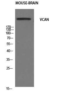
- Western Blot analysis of mouse-brain cells using Versican Polyclonal Antibody. Secondary antibody(catalog#:RS0002) was diluted at 1:20000

- Immunohistochemical analysis of paraffin-embedded human tonsil. 1, Antibody was diluted at 1:200(4° overnight). 2, Tris-EDTA,pH9.0 was used for antigen retrieval. 3,Secondary antibody was diluted at 1:200(room temperature, 30min).


