JAM-B Polyclonal Antibody
- Catalog No.:YT5115
- Applications:WB;IHC;IF;ELISA
- Reactivity:Human;Mouse;Rat
- Target:
- JAM-B
- Fields:
- >>Cell adhesion molecules;>>Tight junction;>>Leukocyte transendothelial migration;>>Epithelial cell signaling in Helicobacter pylori infection
- Gene Name:
- JAM2
- Protein Name:
- Junctional adhesion molecule B
- Human Gene Id:
- 58494
- Human Swiss Prot No:
- P57087
- Mouse Gene Id:
- 67374
- Mouse Swiss Prot No:
- Q9JI59
- Immunogen:
- Synthesized peptide derived from the Internal region of human JAM-B.
- Specificity:
- JAM-B Polyclonal Antibody detects endogenous levels of JAM-B protein.
- Formulation:
- Liquid in PBS containing 50% glycerol, 0.5% BSA and 0.02% sodium azide.
- Source:
- Polyclonal, Rabbit,IgG
- Dilution:
- WB 1:500 - 1:2000. IHC: 1:100-300 ELISA: 1:20000.. IF 1:50-200
- Purification:
- The antibody was affinity-purified from rabbit antiserum by affinity-chromatography using epitope-specific immunogen.
- Concentration:
- 1 mg/ml
- Storage Stability:
- -15°C to -25°C/1 year(Do not lower than -25°C)
- Other Name:
- JAM2;C21orf43;VEJAM;Junctional adhesion molecule B;JAM-B;Junctional adhesion molecule 2;JAM-2;Vascular endothelial junction-associated molecule;VE-JAM;CD322
- Observed Band(KD):
- 33kD
- Background:
- This gene belongs to the immunoglobulin superfamily, and the junctional adhesion molecule (JAM) family. The protein encoded by this gene is a type I membrane protein that is localized at the tight junctions of both epithelial and endothelial cells. It acts as an adhesive ligand for interacting with a variety of immune cell types, and may play a role in lymphocyte homing to secondary lymphoid organs. Alternatively spliced transcript variants have been found for this gene. [provided by RefSeq, Jul 2012],
- Function:
- function:May play a role in the processes of lymphocyte homing to secondary lymphoid organs.,similarity:Belongs to the immunoglobulin superfamily.,similarity:Contains 1 Ig-like C2-type (immunoglobulin-like) domain.,similarity:Contains 1 Ig-like V-type (immunoglobulin-like) domain.,subcellular location:Localized at tight junctions of both epithelial and endothelial cells.,subunit:Interacts with JAM3.,tissue specificity:Highest expression in the heart, placenta, lung, foreskin and lymph node. Prominently expressed on high endothelial venules, also present on the endothelia of other vessels. Localized to the intercellular boundaries of high endothelial cells.,
- Subcellular Location:
- Cell membrane ; Single-pass type I membrane protein . Cell junction . Cell junction, tight junction . Localized at tight junctions of both epithelial and endothelial cells (By similarity). Specifically localized within the somatodendritic compartment of neurons and excluded from the axon (By similarity). .
- Expression:
- Highly expressed in heart, placenta, lung, foreskin and lymph node (PubMed:10779521, PubMed:10945976). Prominently expressed on high endothelial venules and also present on the endothelia of other vessels (at protein level) (PubMed:10779521, PubMed:10945976). Also expressed in the brain in the caudate nuclei (PubMed:31851307).
- June 19-2018
- WESTERN IMMUNOBLOTTING PROTOCOL
- June 19-2018
- IMMUNOHISTOCHEMISTRY-PARAFFIN PROTOCOL
- June 19-2018
- IMMUNOFLUORESCENCE PROTOCOL
- September 08-2020
- FLOW-CYTOMEYRT-PROTOCOL
- May 20-2022
- Cell-Based ELISA│解您多样本WB检测之困扰
- July 13-2018
- CELL-BASED-ELISA-PROTOCOL-FOR-ACETYL-PROTEIN
- July 13-2018
- CELL-BASED-ELISA-PROTOCOL-FOR-PHOSPHO-PROTEIN
- July 13-2018
- Antibody-FAQs
- Products Images

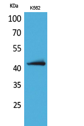
- Western Blot analysis of K562 cells using JAM-B Polyclonal Antibody. Secondary antibody(catalog#:RS0002) was diluted at 1:20000

- Immunohistochemical analysis of paraffin-embedded rat-lung, antibody was diluted at 1:100


