Talin-1 Polyclonal Antibody
- Catalog No.:YT5576
- Applications:WB;IHC;IF;ELISA
- Reactivity:Human;Mouse;Rat
- Target:
- Talin
- Fields:
- >>Rap1 signaling pathway;>>Focal adhesion;>>Platelet activation;>>Shigellosis;>>Human T-cell leukemia virus 1 infection
- Gene Name:
- TLN1
- Protein Name:
- Talin-1
- Human Gene Id:
- 7094
- Human Swiss Prot No:
- Q9Y490
- Mouse Gene Id:
- 21894
- Mouse Swiss Prot No:
- P26039
- Immunogen:
- The antiserum was produced against synthesized peptide derived from the C-terminal region of human TLN1. AA range:2471-2520
- Specificity:
- Talin-1 Polyclonal Antibody detects endogenous levels of Talin-1 protein.
- Formulation:
- Liquid in PBS containing 50% glycerol, 0.5% BSA and 0.02% sodium azide.
- Source:
- Polyclonal, Rabbit,IgG
- Dilution:
- WB 1:500 - 1:2000. IHC: 1:100-300 ELISA: 1:20000. IF 1:100-300 Not yet tested in other applications.
- Purification:
- The antibody was affinity-purified from rabbit antiserum by affinity-chromatography using epitope-specific immunogen.
- Concentration:
- 1 mg/ml
- Storage Stability:
- -15°C to -25°C/1 year(Do not lower than -25°C)
- Other Name:
- TLN1;KIAA1027;TLN;Talin-1
- Observed Band(KD):
- 269kD
- Background:
- This gene encodes a cytoskeletal protein that is concentrated in areas of cell-substratum and cell-cell contacts. The encoded protein plays a significant role in the assembly of actin filaments and in spreading and migration of various cell types, including fibroblasts and osteoclasts. It codistributes with integrins in the cell surface membrane in order to assist in the attachment of adherent cells to extracellular matrices and of lymphocytes to other cells. The N-terminus of this protein contains elements for localization to cell-extracellular matrix junctions. The C-terminus contains binding sites for proteins such as beta-1-integrin, actin, and vinculin. [provided by RefSeq, Feb 2009],
- Function:
- function:Probably involved in connections of major cytoskeletal structures to the plasma membrane. High molecular weight cytoskeletal protein concentrated at regions of cell-substratum contact and, in lymphocytes, at cell-cell contacts.,similarity:Contains 1 FERM domain.,similarity:Contains 1 I/LWEQ domain.,subcellular location:Colocalizes with LAYN at the membrane ruffles.,subunit:Binds with high affinity to vinculin and with low affinity to integrins. Interacts with PIP5K1C and NRAP. Interacts with LAYN. Interacts with SYNM.,
- Subcellular Location:
- Cell projection, ruffle membrane ; Peripheral membrane protein ; Cytoplasmic side . Cytoplasm, cytoskeleton . Cell surface . Cell junction, focal adhesion . Colocalizes with LAYN at the membrane ruffles. Localized preferentially in focal adhesions than fibrillar adhesions (By similarity). .
- Expression:
- Aorta,Brain,Embryonic carcinoma,Platelet,
- June 19-2018
- WESTERN IMMUNOBLOTTING PROTOCOL
- June 19-2018
- IMMUNOHISTOCHEMISTRY-PARAFFIN PROTOCOL
- June 19-2018
- IMMUNOFLUORESCENCE PROTOCOL
- September 08-2020
- FLOW-CYTOMEYRT-PROTOCOL
- May 20-2022
- Cell-Based ELISA│解您多样本WB检测之困扰
- July 13-2018
- CELL-BASED-ELISA-PROTOCOL-FOR-ACETYL-PROTEIN
- July 13-2018
- CELL-BASED-ELISA-PROTOCOL-FOR-PHOSPHO-PROTEIN
- July 13-2018
- Antibody-FAQs
- Products Images
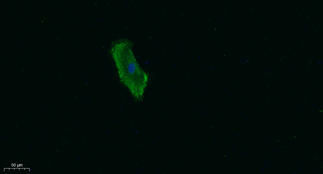
- Immunofluorescence analysis of A549. 1,primary Antibody was diluted at 1:200(4°C overnight). 2, Goat Anti Rabbit IgG (H&L) - Alexa Fluor 488 Secondary antibody was diluted at 1:1000(room temperature, 50min).3, Picture B: DAPI(blue) 10min.
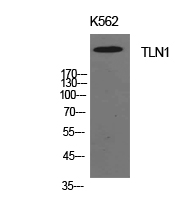
- Western Blot analysis of K562 cells using Talin-1 Polyclonal Antibody. Antibody was diluted at 1:1000. Secondary antibody(catalog#:RS0002) was diluted at 1:20000

- Immunohistochemical analysis of paraffin-embedded human-kidney, antibody was diluted at 1:100
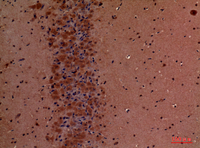
- Immunohistochemical analysis of paraffin-embedded mouse-brain, antibody was diluted at 1:100


