CD6 Polyclonal Antibody
- Catalog No.:YT5577
- Applications:WB;IHC;IF;ELISA
- Reactivity:Human;Mouse
- Target:
- CD6
- Fields:
- >>Cell adhesion molecules
- Gene Name:
- CD6
- Protein Name:
- T-cell differentiation antigen CD6
- Human Gene Id:
- 923
- Human Swiss Prot No:
- P30203
- Mouse Swiss Prot No:
- Q61003
- Immunogen:
- The antiserum was produced against synthesized peptide derived from the Internal region of human CD6. AA range:361-410
- Specificity:
- CD6 Polyclonal Antibody detects endogenous levels of CD6 protein.
- Formulation:
- Liquid in PBS containing 50% glycerol, 0.5% BSA and 0.02% sodium azide.
- Source:
- Polyclonal, Rabbit,IgG
- Dilution:
- WB 1:500 - 1:2000. IHC: 1:100-1:300. ELISA: 1:10000.. IF 1:50-200
- Purification:
- The antibody was affinity-purified from rabbit antiserum by affinity-chromatography using epitope-specific immunogen.
- Concentration:
- 1 mg/ml
- Storage Stability:
- -15°C to -25°C/1 year(Do not lower than -25°C)
- Other Name:
- CD6;T-cell differentiation antigen CD6;T12;TP120;CD6
- Observed Band(KD):
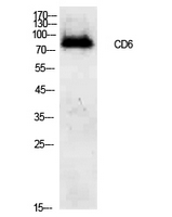
- 73kD
- Background:
- This gene encodes a protein found on the outer membrane of T-lymphocytes as well as some other immune cells. The encoded protein contains three scavenger receptor cysteine-rich (SRCR) domains and a binding site for an activated leukocyte cell adhesion molecule. The gene product is important for continuation of T cell activation. This gene may be associated with susceptibility to multiple sclerosis (PMID: 19525953, 21849685). Multiple transcript variants encoding different isoforms have been found for this gene. [provided by RefSeq, Dec 2011],
- Function:
- function:Involved in cell adhesion. Binds to CD166.,PTM:After T-cell activation, becomes hyperphosphorylated on Ser and Thr residues and phosphorylated on Tyr residues.,PTM:Contains intrachain disulfide bond(s).,similarity:Contains 3 SRCR domains.,tissue specificity:Expressed by thymocytes, mature T-cells, a subset of B-cells known as B-1 cells, and by some cells in the brain.,
- Subcellular Location:
- Cell membrane ; Single-pass type I membrane protein . Detected at the immunological synapse, i.e, at the contact zone between antigen-presenting dendritic cells and T-cells (PubMed:15294938, PubMed:16352806). Colocalizes with the TCR/CD3 complex at the immunological synapse (PubMed:15294938). .; [Soluble CD6]: Secreted . The origins of the secreted form are not clear, but it might be created by proteolytic shedding of the ectodomain. .
- Expression:
- Detected on thymocytes (PubMed:15294938). Detected on peripheral blood T-cells (PubMed:15048703, PubMed:16352806). Detected on natural killer (NK) cells (PubMed:16352806). Soluble CD6 is detected in blood serum (at protein level) (PubMed:17601777). Detected in spleen, thymus, appendix, lymph node and peripheral blood leukocytes (PubMed:9013954). Expressed by thymocytes, mature T-cells, a subset of B-cells known as B-1 cells, and by some cells in the brain.
- June 19-2018
- WESTERN IMMUNOBLOTTING PROTOCOL
- June 19-2018
- IMMUNOHISTOCHEMISTRY-PARAFFIN PROTOCOL
- June 19-2018
- IMMUNOFLUORESCENCE PROTOCOL
- September 08-2020
- FLOW-CYTOMEYRT-PROTOCOL
- May 20-2022
- Cell-Based ELISA│解您多样本WB检测之困扰
- July 13-2018
- CELL-BASED-ELISA-PROTOCOL-FOR-ACETYL-PROTEIN
- July 13-2018
- CELL-BASED-ELISA-PROTOCOL-FOR-PHOSPHO-PROTEIN
- July 13-2018
- Antibody-FAQs
- Products Images

- Western Blot analysis of 293 cells using CD6 Polyclonal Antibody. Secondary antibody(catalog#:RS0002) was diluted at 1:20000

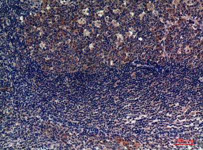
- Immunohistochemical analysis of paraffin-embedded human-tonsils, antibody was diluted at 1:100


