- 靶点:
- Tubulin β
- 简介:
- >>Phagosome;>>Gap junction;>>Alzheimer disease;>>Parkinson disease;>>Amyotrophic lateral sclerosis;>>Huntington disease;>>Prion disease;>>Pathways of neurodegeneration - multiple diseases;>>Pathogenic Escherichia coli infection;>>Salmonella infection
- 基因名称:
- TUBB1
- 蛋白名称:
- Tubulin beta-1 chain
- Human Gene Id:
- 81027
- Human Swiss Prot No:
- Q9H4B7
- Mouse Gene Id:
- 545486
- Mouse Swiss Prot No:
- A2AQ07
- 免疫原:
- Synthetic Peptide of β I tubulin
- 特异性:
- The antibody detects endgenousβ I tubulin proteins.
- 组成:
- PBS, pH 7.4, containing 0.5%BSA, 0.02% sodium azide as Preservative and 50% Glycerol.
- 来源:
- Monoclonal, Mouse
- 稀释:
- WB 1:500-10000 IF 1:200 IHC 1:50-300
- 纯化工艺:
- The antibody was affinity-purified from mouse ascites by affinity-chromatography using specific immunogen.
- 储存:
- -15°C to -25°C/1 year(Do not lower than -25°C)
- 其他名称:
- TUBB1;Tubulin beta-1 chain;TUBB2A;TUBB2;Tubulin beta-2A chain;TUBB2B;Tubulin beta-2B chain;TUBB4B;TUBB2C;Tubulin beta-4B chain;Tubulin beta-2 chain;Tubulin beta-2C chain;TUBB3;TUBB4;Tubulin beta-3 chain;Tubulin beta-4 chain;Tubulin beta-III;TUBB4A;TUBB4;TUBB5;Tubulin beta-4A chain;Tubulin 5 beta;Tubulin beta-4 chain;TUBB;TUBB5;OK/SW-cl.56;Tubulin beta chain;Tubulin beta-5 chain;TUBB6;Tubulin beta-6 chain;TUBB8;Tubulin beta-8 chain
- 实测条带:
- 50kD
- 背景:
- This gene encodes a member of the beta tubulin protein family. Beta tubulins are one of two core protein families (alpha and beta tubulins) that heterodimerize and assemble to form microtubules. This protein is specifically expressed in platelets and megakaryocytes and may be involved in proplatelet production and platelet release. A mutations in this gene is associated with autosomal dominant macrothrombocytopenia. Two pseudogenes of this gene are found on chromosome Y.[provided by RefSeq, Jul 2010],
- 功能:
- function:Tubulin is the major constituent of microtubules. It binds two moles of GTP, one at an exchangeable site on the beta chain and one at a non-exchangeable site on the alpha-chain.,similarity:Belongs to the tubulin family.,subunit:Dimer of alpha and beta chains.,
- 细胞定位:
- Cytoplasm, cytoskeleton .
- 组织表达:
- Hematopoietic cell-specific. Major isotype in leukocytes, where it represents 50% of all beta-tubulins.
Protease-activated receptor-2 regulates glial scar formation via JNK signaling.. PHYSIOLOGICAL RESEARCH Physiol Res. 2019 Jan;68(2):305-316 IF Rat 1:400 spinal cord
货号:YM3037
- June 19-2018
- WESTERN IMMUNOBLOTTING PROTOCOL
- June 19-2018
- IMMUNOHISTOCHEMISTRY-PARAFFIN PROTOCOL
- June 19-2018
- IMMUNOFLUORESCENCE PROTOCOL
- September 08-2020
- FLOW-CYTOMEYRT-PROTOCOL
- May 20-2022
- Cell-Based ELISA│解您多样本WB检测之困扰
- July 13-2018
- CELL-BASED-ELISA-PROTOCOL-FOR-ACETYL-PROTEIN
- July 13-2018
- CELL-BASED-ELISA-PROTOCOL-FOR-PHOSPHO-PROTEIN
- July 13-2018
- Antibody-FAQs
- 产品图片
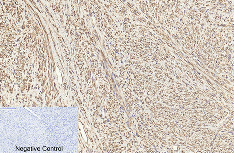
- Immunohistochemical analysis of paraffin-embedded Human-uterus-cancer tissue. 1,β I tubulin Monoclonal Antibody(3F7) was diluted at 1:200(4°C,overnight). 2, Sodium citrate pH 6.0 was used for antibody retrieval(>98°C,20min). 3,Secondary antibody was diluted at 1:200(room tempeRature, 30min). Negative control was used by secondary antibody only.
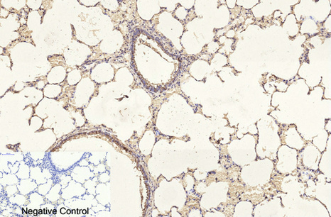
- Immunohistochemical analysis of paraffin-embedded Rat-lung tissue. 1,β I tubulin Monoclonal Antibody(3F7) was diluted at 1:200(4°C,overnight). 2, Sodium citrate pH 6.0 was used for antibody retrieval(>98°C,20min). 3,Secondary antibody was diluted at 1:200(room tempeRature, 30min). Negative control was used by secondary antibody only.
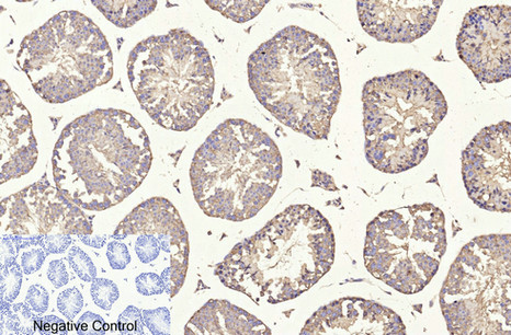
- Immunohistochemical analysis of paraffin-embedded Mouse-testis tissue. 1,β I tubulin Monoclonal Antibody(3F7) was diluted at 1:200(4°C,overnight). 2, Sodium citrate pH 6.0 was used for antibody retrieval(>98°C,20min). 3,Secondary antibody was diluted at 1:200(room tempeRature, 30min). Negative control was used by secondary antibody only.
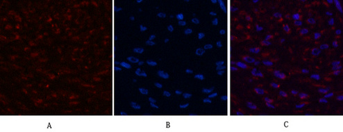
- Immunofluorescence analysis of Human-breast-cancer tissue. 1,β I tubulin Monoclonal Antibody(3F7)(red) was diluted at 1:200(4°C,overnight). 2, Cy3 labled Secondary antibody was diluted at 1:300(room temperature, 50min).3, Picture B: DAPI(blue) 10min. Picture A:Target. Picture B: DAPI. Picture C: merge of A+B

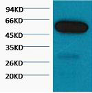
- Western blot analysis of 1) Hela, 2) Mouse Brain Tissue, 3) Rat Brain Tissue, diluted at 1:5000.