Vimentin (ABT281) IHC kit
- Catalog No.:IHCM6918
- Applications:IHC
- Reactivity:Human;Mouse;Rat;
- Target:
- Vimentin
- Fields:
- >>Epstein-Barr virus infection;>>MicroRNAs in cancer
- Gene Name:
- VIM
- Protein Name:
- Vimentin
- Human Gene Id:
- 7431
- Human Swiss Prot No:
- P08670
- Immunogen:
- Synthesized peptide derived from human Vimentin AA range: 400-466
- Specificity:
- The antibody can specifically recognize human Vimentin protein.
- Source:
- Mouse, Monoclonal/IgG1, kappa
- Purification:
- The antibody was affinity-purified from ascites by affinity-chromatography using specific immunogen.
- Storage Stability:
- 2°C to 8°C/1 year
- Other Name:
- Vimentin
- Background:
- This gene encodes a member of the intermediate filament family. Intermediate filamentents, along with microtubules and actin microfilaments, make up the cytoskeleton. The protein encoded by this gene is responsible for maintaining cell shape, integrity of the cytoplasm, and stabilizing cytoskeletal interactions. It is also involved in the immune response, and controls the transport of low-density lipoprotein (LDL)-derived cholesterol from a lysosome to the site of esterification. It functions as an organizer of a number of critical proteins involved in attachment, migration, and cell signaling. Mutations in this gene causes a dominant, pulverulent cataract.[provided by RefSeq, Jun 2009],
- Function:
- function:Vimentins are class-III intermediate filaments found in various non-epithelial cells, especially mesenchymal cells.,online information:Vimentin entry,PTM:One of the most prominent phosphoproteins in various cells of mesenchymal origin. Phosphorylation is enhanced during cell division, at which time vimentin filaments are significantly reorganized.,sequence caution:Intron retention.,similarity:Belongs to the intermediate filament family.,subunit:Homopolymer. Interacts with HCV core protein. Interacts with LGSN and SYNM.,tissue specificity:Highly expressed in fibroblasts, some expression in T- and B-lymphocytes, and little or no expression in Burkitt's lymphoma cell lines. Expressed in many hormone-independent mammary carcinoma cell lines.,
- Subcellular Location:
- Cytoplasmic
- Expression:
- Highly expressed in fibroblasts, some expression in T- and B-lymphocytes, and little or no expression in Burkitt's lymphoma cell lines. Expressed in many hormone-independent mammary carcinoma cell lines.
- June 19-2018
- WESTERN IMMUNOBLOTTING PROTOCOL
- June 19-2018
- IMMUNOHISTOCHEMISTRY-PARAFFIN PROTOCOL
- June 19-2018
- IMMUNOFLUORESCENCE PROTOCOL
- September 08-2020
- FLOW-CYTOMEYRT-PROTOCOL
- May 20-2022
- Cell-Based ELISA│解您多样本WB检测之困扰
- July 13-2018
- CELL-BASED-ELISA-PROTOCOL-FOR-ACETYL-PROTEIN
- July 13-2018
- CELL-BASED-ELISA-PROTOCOL-FOR-PHOSPHO-PROTEIN
- July 13-2018
- Antibody-FAQs
- Products Images
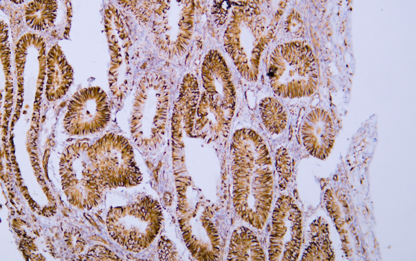
- Human endometrial adenocarcinoma tissue was stained with Anti-Vimentin (ABT281) Antibody
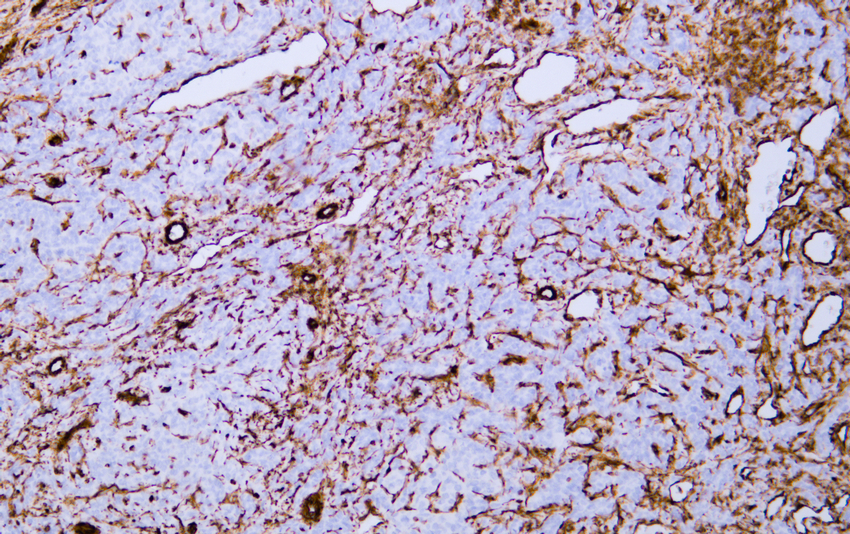
- Human hepatocellular carcinoma tissue was stained with Anti-Vimentin (ABT281) Antibody
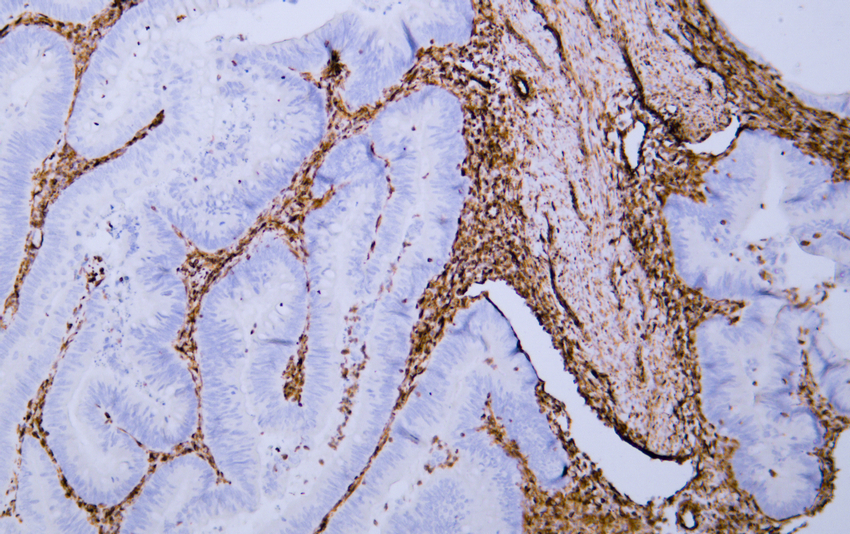
- Human rectal carcinoma tissue was stained with Anti-Vimentin (ABT281) Antibody
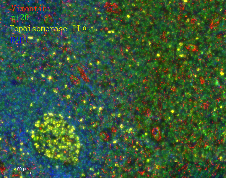
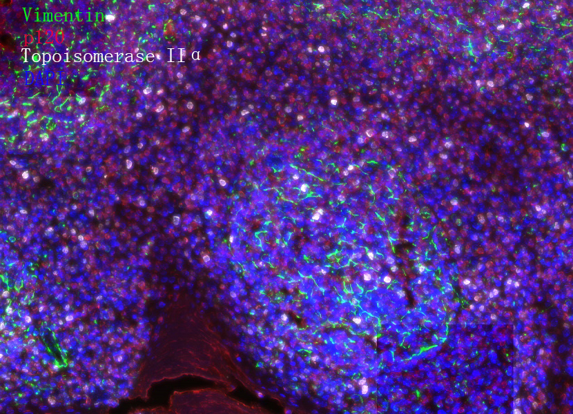
- Fluorescence multiplex immunohistochemical analysis of Human tonsil tissue (formalin-fixed paraffin-embedded section). Merged staining of Anti-Vimentin (YM6918), Anti-p120 (YM6086), Anti-Topoisomerase IIα (YM6914). The immunostaining was performed on a Leica Biosystems BOND® MAX instrument with an Sextuple-Fluorescence kit (RS0039, Immunoway). The section was incubated in 3 rounds of staining; sequentially for Anti-Vimentin (YM6918 1:200), Anti-p120 (YM6086 1:200), Anti-Topoisomerase IIα (YM6914 1:200).; each using a separate fluorescent tyramide signal amplification system. EDTA based antigen retrieval (Leica Biosystems BOND® Epitope Retrieval Solution 2, pH 9.0, 20 minutes) was used in between rounds of tyramide signal amplification to remove the antibody from the previous round, to avoid any cross-reactivity. DAPI (dark blue) was used as a nuclear counter stain. Microscopy and pseudocoloring of individual dyes was performed using a Slideviewer Imaging System (3D histech).
- Fluorescence multiplex immunohistochemical analysis of Human tonsil tissue (formalin-fixed paraffin-embedded section). Merged staining of Anti-Vimentin (YM6918), Anti-p120 (YM6086), Anti-Topoisomerase IIα (YM6914). The immunostaining was performed on a Leica Biosystems BOND® MAX instrument with an Sextuple-Fluorescence kit (RS0039, Immunoway). The section was incubated in 3 rounds of staining; sequentially for Anti-Vimentin (YM6918 1:200), Anti-p120 (YM6086 1:200), Anti-Topoisomerase IIα (YM6914 1:200).; each using a separate fluorescent tyramide signal amplification system. EDTA based antigen retrieval (Leica Biosystems BOND® Epitope Retrieval Solution 2, pH 9.0, 20 minutes) was used in between rounds of tyramide signal amplification to remove the antibody from the previous round, to avoid any cross-reactivity. DAPI (dark blue) was used as a nuclear counter stain. Microscopy and pseudocoloring of individual dyes was performed using a Slideviewer Imaging System (3D histech).