PCNA Monoclonal Antibody(12D10)
- Catalog No.:YM3031
- Applications:WB;IF;IHC
- Reactivity:Human;Rat;Mouse
- Target:
- PCNA
- Fields:
- >>DNA replication;>>Base excision repair;>>Nucleotide excision repair;>>Mismatch repair;>>Cell cycle;>>Tight junction;>>Hepatitis B
- Gene Name:
- PCNA
- Protein Name:
- Proliferating cell nuclear antigen
- Human Gene Id:
- 5111
- Human Swiss Prot No:
- P12004
- Mouse Gene Id:
- 18538
- Mouse Swiss Prot No:
- P17918
- Rat Gene Id:
- 25737
- Rat Swiss Prot No:
- P04961
- Immunogen:
- Synthetic Peptide of PCNA
- Specificity:
- The antibody detects endogenous PCNA protein.
- Formulation:
- PBS, pH 7.4, containing 0.5%BSA, 0.02% sodium azide as Preservative and 50% Glycerol.
- Source:
- Monoclonal, Mouse
- Dilution:
- WB 1:5000 IHC 1:200 IF 1:200
- Purification:
- The antibody was affinity-purified from mouse ascites by affinity-chromatography using specific immunogen.
- Storage Stability:
- -15°C to -25°C/1 year(Do not lower than -25°C)
- Other Name:
- PCNA;Proliferating cell nuclear antigen;PCNA;Cyclin
- Observed Band(KD):
- 30-33kd
- Background:
- The protein encoded by this gene is found in the nucleus and is a cofactor of DNA polymerase delta. The encoded protein acts as a homotrimer and helps increase the processivity of leading strand synthesis during DNA replication. In response to DNA damage, this protein is ubiquitinated and is involved in the RAD6-dependent DNA repair pathway. Two transcript variants encoding the same protein have been found for this gene. Pseudogenes of this gene have been described on chromosome 4 and on the X chromosome. [provided by RefSeq, Jul 2008],
- Function:
- disease:Antibodies are present in sera from patients with systemic lupus erythematosus.,function:This protein is an auxiliary protein of DNA polymerase delta and is involved in the control of eukaryotic DNA replication by increasing the polymerase's processibility during elongation of the leading strand.,online information:PCNA entry,PTM:Upon methyl methanesulfonate-induced DNA damage, mono-ubiquitinated by the UBE2B-RAD18 complex on Lys-164. This induces non-canonical poly-ubiquitination on Lys-164 through 'Lys-63' linkage of ubiquitin moieties by the E2 complex UBE2N-UBE2V2 and the E3 ligases RNF8 and SHPRH, which are required for DNA repair.,similarity:Belongs to the PCNA family.,subunit:Homotrimer. Interacts with KCTD10. Interacts with PPP1R15A (By similarity). Forms a complex with activator 1 heteropentamer in the presence of ATP. Interacts with POLH, POLK, DNMT1, ERCC5/XPG, FEN1, C
- Subcellular Location:
- Nucleus . Colocalizes with CREBBP, EP300 and POLD1 to sites of DNA damage (PubMed:24939902). Forms nuclear foci representing sites of ongoing DNA replication and vary in morphology and number during S phase (PubMed:15543136). Co-localizes with SMARCA5/SNF2H and BAZ1B/WSTF at replication foci during S phase (PubMed:15543136). Together with APEX2, is redistributed in discrete nuclear foci in presence of oxidative DNA damaging agents. .
- Expression:
- Bone marrow,Fetal brain cortex,Lung,Placenta,
Inhibition of the Ras/ERK1/2 pathway contributes to the protective effect of ginsenoside Re against intimal hyperplasia. Food & Function Food Funct. 2021 May;: IF Rat 1 : 200 Carotid arteries
CD137–CD137L interaction modulates neointima formation and the phenotype transformation of vascular smooth muscle cells via NFATc1 signaling. MOLECULAR AND CELLULAR BIOCHEMISTRY Mol Cell Biochem. 2018 Feb;439(1):65-74 WB Mouse VSMCs
Overexpression of miR‐200b inhibits the cell proliferation and promotes apoptosis of human hypertrophic scar fibroblasts in vitro. JOURNAL OF DERMATOLOGY WB Human hHSFs cell
High mobility group protein B1 (HMGB1) interacts with receptor for advanced glycation end products (RAGE) to promote airway smooth muscle cell proliferation through ERK and NF-κB pathways. International Journal of Clinical and Experimental Pathology Int J Clin Exp Patho. 2019; 12(9): 3268–3278 WB Rat RASMCs
Huaier Restrains Proliferative and Migratory Potential of Hepatocellular Carcinoma Cells Partially Through Decreased Yes-Associated Protein 1. Journal of Cancer J Cancer. 2017; 8(19): 4087–4097 WB Human Bel-7404 cell,SMMC-7721 cell
Gao, Chenying, et al. "Inhibition of Ras/ERK1/2 pathway contributes to the protective effect of Ginsenoside Re against intimal hyperplasia." Food & Function (2021).
PFO5DoDA disrupts hepatic homeostasis primarily through glucocorticoid signaling inhibition JOURNAL OF HAZARDOUS MATERIALS Xinyu Liu WB Mouse liver
- June 19-2018
- WESTERN IMMUNOBLOTTING PROTOCOL
- June 19-2018
- IMMUNOHISTOCHEMISTRY-PARAFFIN PROTOCOL
- June 19-2018
- IMMUNOFLUORESCENCE PROTOCOL
- September 08-2020
- FLOW-CYTOMEYRT-PROTOCOL
- May 20-2022
- Cell-Based ELISA│解您多样本WB检测之困扰
- July 13-2018
- CELL-BASED-ELISA-PROTOCOL-FOR-ACETYL-PROTEIN
- July 13-2018
- CELL-BASED-ELISA-PROTOCOL-FOR-PHOSPHO-PROTEIN
- July 13-2018
- Antibody-FAQs
- Products Images

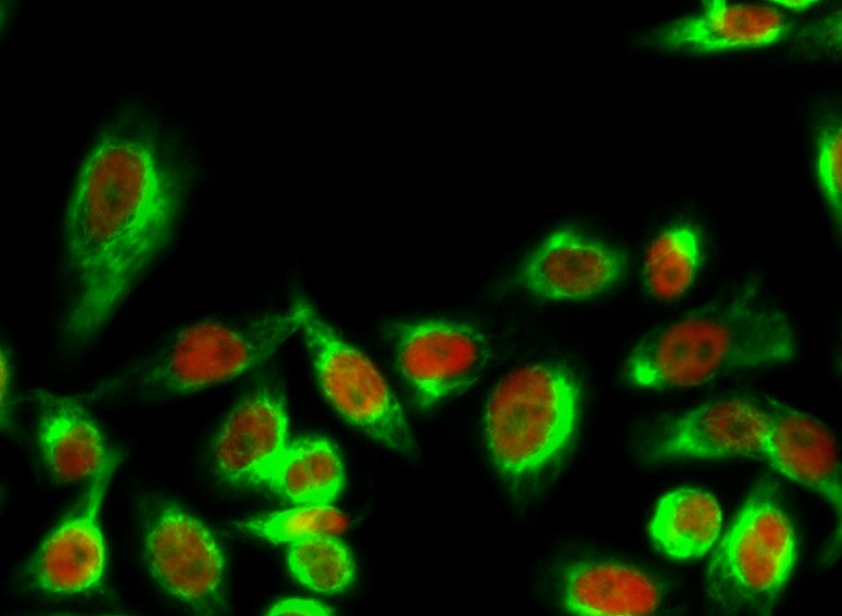
- Immunofluorescence analysis of Hela cell. 1,14-3-3 θ/τ (phospho Ser232) Polyclonal Antibody(green) was diluted at 1:200(4° overnight). (red) was diluted at 1:200(4° overnight). 2, Goat Anti Rabbit Alexa Fluor 488 Catalog:RS3211 was diluted at 1:1000(room temperature, 50min). Goat Anti Mouse Alexa Fluor 594 Catalog:RS3608 was diluted at 1:1000(room temperature, 50min).

- Immunofluorescence analysis of Human-lung-cancer tissue. 1,PCNA Monoclonal Antibody(12D10)(red) was diluted at 1:200(4°C,overnight). 2, Cy3 labled Secondary antibody was diluted at 1:300(room temperature, 50min).3, Picture B: DAPI(blue) 10min. Picture A:Target. Picture B: DAPI. Picture C: merge of A+B

- Immunofluorescence analysis of Rat-testis tissue. 1,PCNA Monoclonal Antibody(12D10)(red) was diluted at 1:200(4°C,overnight). 2, Cy3 labled Secondary antibody was diluted at 1:300(room temperature, 50min).3, Picture B: DAPI(blue) 10min. Picture A:Target. Picture B: DAPI. Picture C: merge of A+B

- Various whole cell lysates were separated by 10% SDS-PAGE, and the membrane was blotted with anti-PCNA antibody. The HRP-conjugated anti-Mouse IgG antibody was used to detect the antibody.
.jpg)
- Human colon tissue was stained with Anti-Proliferating Cell Nuclear Antigen (ABT221) Antibody
.jpg)
- Human colon tissue was stained with Anti-Proliferating Cell Nuclear Antigen (ABT221) Antibody
.jpg)
- Human colon tissue was stained with Anti-Proliferating Cell Nuclear Antigen (ABT221) Antibody
