Smad3 (phospho Ser425) Polyclonal Antibody
- Catalog No.:YP0585
- Applications:IF;WB;IHC;ELISA
- Reactivity:Human;Mouse;Rat
- Target:
- Smad3
- Fields:
- >>FoxO signaling pathway;>>Cell cycle;>>Endocytosis;>>Cellular senescence;>>Wnt signaling pathway;>>TGF-beta signaling pathway;>>Apelin signaling pathway;>>Hippo signaling pathway;>>Adherens junction;>>Signaling pathways regulating pluripotency of stem cells;>>Th17 cell differentiation;>>AGE-RAGE signaling pathway in diabetic complications;>>Hepatitis B;>>Human T-cell leukemia virus 1 infection;>>Pathways in cancer;>>Colorectal cancer;>>Pancreatic cancer;>>Chronic myeloid leukemia;>>Hepatocellular carcinoma;>>Gastric cancer;>>Inflammatory bowel disease;>>Diabetic cardiomyopathy
- Gene Name:
- SMAD3
- Protein Name:
- Mothers against decapentaplegic homolog 3
- Human Gene Id:
- 4088
- Human Swiss Prot No:
- P84022
- Mouse Gene Id:
- 17127
- Mouse Swiss Prot No:
- Q8BUN5
- Rat Gene Id:
- 25631
- Rat Swiss Prot No:
- P84025
- Immunogen:
- The antiserum was produced against synthesized peptide derived from human Smad3 around the phosphorylation site of Ser425. AA range:376-425
- Specificity:
- Phospho-Smad3 (S425) Polyclonal Antibody detects endogenous levels of Smad3 protein only when phosphorylated at S425.
- Formulation:
- Liquid in PBS containing 50% glycerol, 0.5% BSA and 0.02% sodium azide.
- Source:
- Polyclonal, Rabbit,IgG
- Dilution:
- IF 1:50-200 WB 1:500 - 1:2000. IHC 1:100 - 1:300. ELISA: 1:40000. Not yet tested in other applications.
- Purification:
- The antibody was affinity-purified from rabbit antiserum by affinity-chromatography using epitope-specific immunogen.
- Concentration:
- 1 mg/ml
- Storage Stability:
- -15°C to -25°C/1 year(Do not lower than -25°C)
- Other Name:
- SMAD3;MADH3;Mothers against decapentaplegic homolog 3;MAD homolog 3;Mad3;Mothers against DPP homolog 3;hMAD-3;JV15-2;SMAD family member 3;SMAD 3;Smad3;hSMAD3
- Observed Band(KD):
- 58kD
- Background:
- The protein encoded by this gene belongs to the SMAD, a family of proteins similar to the gene products of the Drosophila gene 'mothers against decapentaplegic' (Mad) and the C. elegans gene Sma. SMAD proteins are signal transducers and transcriptional modulators that mediate multiple signaling pathways. This protein functions as a transcriptional modulator activated by transforming growth factor-beta and is thought to play a role in the regulation of carcinogenesis. [provided by RefSeq, Apr 2009],
- Function:
- disease:Defects in SMAD3 may be a cause of colorectal cancer (CRC) [MIM:114500].,domain:The MH2 domain is sufficient to carry protein nuclear export.,function:Transcriptional modulator activated by TGF-beta (transforming growth factor) and activin type 1 receptor kinase. SMAD3 is a receptor-regulated SMAD (R-SMAD).,PTM:Phosphorylated on serine by TGF-beta and activin type 1 receptor kinases.,similarity:Belongs to the dwarfin/SMAD family.,similarity:Contains 1 MH1 (MAD homology 1) domain.,similarity:Contains 1 MH2 (MAD homology 2) domain.,subcellular location:In the cytoplasm in the absence of ligand. Migration to the nucleus when complexed with Smad4.,subunit:Interacts with HGS. Interacts with NEDD4L in response to TGF-beta. Interacts with TTRAP (By similarity). Interacts with SARA (SMAD anchor for receptor activation); form trimers with another SMAD3 and the co-SMAD SMAD4. Interacts wit
- Subcellular Location:
- Cytoplasm . Nucleus . Cytoplasmic and nuclear in the absence of TGF-beta. On TGF-beta stimulation, migrates to the nucleus when complexed with SMAD4 (PubMed:15799969, PubMed:21145499). Through the action of the phosphatase PPM1A, released from the SMAD2/SMAD4 complex, and exported out of the nucleus by interaction with RANBP1 (PubMed:16751101, PubMed:19289081). Co-localizes with LEMD3 at the nucleus inner membrane (PubMed:15601644). MAPK-mediated phosphorylation appears to have no effect on nuclear import (PubMed:19218245). PDPK1 prevents its nuclear translocation in response to TGF-beta (PubMed:17327236). Localized mainly to the nucleus in the early stages of embryo development with expression becoming evident in the cytoplasm of the inner cell mass at the blastocyst stage (By similarity)
- Expression:
- Brain,Colon carcinoma,Esophagus tumor,Pancreas,Placenta,Spleen,Umbilical cord blood
IL-37 suppresses hepatocellular carcinoma growth by converting pSmad3 signaling from JNK/pSmad3L/c-Myc oncogenic signaling to pSmad3C/P21 tumor-suppressive signaling. Oncotarget Oncotarget. 2016 Dec 20; 7(51): 85079–85096 WB Human SMMC-7721 cell
Honokiol-mesoporous Silica Nanoparticles Inhibit Vascular Restenosis via the Suppression of TGF-β Signaling Pathway. International Journal of Nanomedicine Int J Nanomed. 2020; 15: 5239–5252 WB Rat Vascular smooth muscle cells (VSMCs)
Inhibitory effect of tranilast on the myofibroblast differentiation of rat mesenchymal stem cells induced by transforming growth factor‑β1 in vitro. Molecular Medicine Reports Mol Med Rep. 2018 Dec;18(6):5693-5700 WB Rat 1:500 MSCs
PDGFBB improved the biological function of menstrual blood-derived stromal cells and the anti-fibrotic properties of exosomes Stem Cell Research & Therapy Jichun Tan IF,WB Human endometrial stromal cells (EndoSCs)
The circadian clock gene, BMAL1, promotes radiosensitization in nasopharyngeal carcinoma by inhibiting the epithelial-to-mesenchymal transition via the TGF-β1/Smads/Snail1 axis ORAL ONCOLOGY Yuxin Li WB Human 1:2000 Nasopharyngeal carcinoma (NPC) 5-8F cell
- June 19-2018
- WESTERN IMMUNOBLOTTING PROTOCOL
- June 19-2018
- IMMUNOHISTOCHEMISTRY-PARAFFIN PROTOCOL
- June 19-2018
- IMMUNOFLUORESCENCE PROTOCOL
- September 08-2020
- FLOW-CYTOMEYRT-PROTOCOL
- May 20-2022
- Cell-Based ELISA│解您多样本WB检测之困扰
- July 13-2018
- CELL-BASED-ELISA-PROTOCOL-FOR-ACETYL-PROTEIN
- July 13-2018
- CELL-BASED-ELISA-PROTOCOL-FOR-PHOSPHO-PROTEIN
- July 13-2018
- Antibody-FAQs
- Products Images
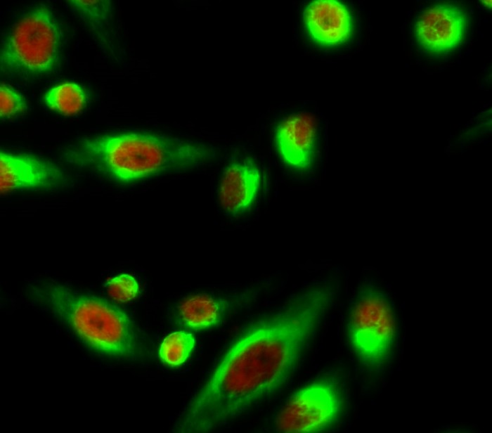
- Immunofluorescence analysis of Hela cell. 1,Smad3 (phospho Ser425) Polyclonal Antibody(red) was diluted at 1:200(4° overnight). ATG5 mouse Monoclonal Antibody(3C7)(green) was diluted at 1:200(4° overnight). 2, Goat Anti Rabbit Alexa Fluor 594 Catalog:RS3611 was diluted at 1:1000(room temperature, 50min). Goat Anti Mouse Alexa Fluor 488 Catalog:RS3208 was diluted at 1:1000(room temperature, 50min).
-if-119.jpg)
- Immunofluorescence analysis of rat-lung tissue. 1,Smad3 (phospho Ser425) Polyclonal Antibody(red) was diluted at 1:200(4°C,overnight). 2, Cy3 labled Secondary antibody was diluted at 1:300(room temperature, 50min).3, Picture B: DAPI(blue) 10min. Picture A:Target. Picture B: DAPI. Picture C: merge of A+B
-if-120.jpg)
- Immunofluorescence analysis of rat-lung tissue. 1,Smad3 (phospho Ser425) Polyclonal Antibody(red) was diluted at 1:200(4°C,overnight). 2, Cy3 labled Secondary antibody was diluted at 1:300(room temperature, 50min).3, Picture B: DAPI(blue) 10min. Picture A:Target. Picture B: DAPI. Picture C: merge of A+B
-if-121.jpg)
- Immunofluorescence analysis of mouse-kidney tissue. 1,Smad3 (phospho Ser425) Polyclonal Antibody(red) was diluted at 1:200(4°C,overnight). 2, Cy3 labled Secondary antibody was diluted at 1:300(room temperature, 50min).3, Picture B: DAPI(blue) 10min. Picture A:Target. Picture B: DAPI. Picture C: merge of A+B
-if-122.jpg)
- Immunofluorescence analysis of mouse-kidney tissue. 1,Smad3 (phospho Ser425) Polyclonal Antibody(red) was diluted at 1:200(4°C,overnight). 2, Cy3 labled Secondary antibody was diluted at 1:300(room temperature, 50min).3, Picture B: DAPI(blue) 10min. Picture A:Target. Picture B: DAPI. Picture C: merge of A+B
poly-ihc-mouse-lung.jpg)
- Immunohistochemical analysis of paraffin-embedded Mouse-lung tissue. 1,Smad3 (phospho Ser425) Polyclonal Antibody was diluted at 1:200(4°C,overnight). 2, Sodium citrate pH 6.0 was used for antibody retrieval(>98°C,20min). 3,Secondary antibody was diluted at 1:200(room tempeRature, 30min). Negative control was used by secondary antibody only.
- Western Blot analysis of various cells using Phospho-Smad3 (S425) Polyclonal Antibody diluted at 1:500
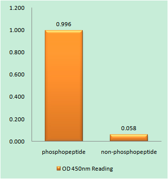
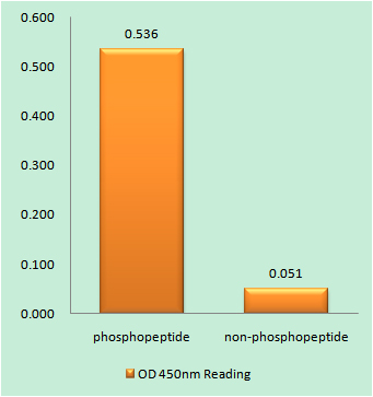
- Enzyme-Linked Immunosorbent Assay (Phospho-ELISA) for Immunogen Phosphopeptide (Phospho-left) and Non-Phosphopeptide (Phospho-right), using Smad3 (Phospho-Ser425) Antibody
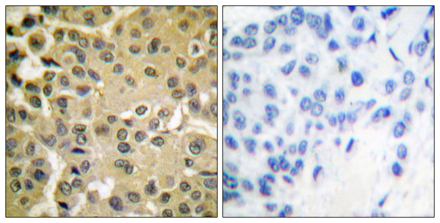
- Immunohistochemistry analysis of paraffin-embedded human breast carcinoma, using Smad3 (Phospho-Ser425) Antibody. The picture on the right is blocked with the phospho peptide.
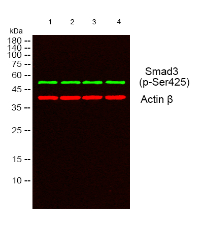
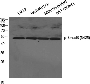
- Western blot analysis of lysates from 1) L929, 2) RAT MUSLE,3)MOUSE-BRAIN, 4) RAT- KIDNEY cells, (Green) primary antibody was diluted at 1:1000, 4°over night, secondary antibody(cat:RS23920)was diluted at 1:10000, 37° 1hour. (Red) Actin β Monoclonal Antibody(5B7) (cat:YM3028) antibody was diluted at 1:5000 as loading control, 4° over night,secondary antibody(cat:RS23710)was diluted at 1:10000, 37° 1hour.