Septin 1 Polyclonal Antibody
- Catalog No.:YT4245
- Applications:WB;IHC;IF;ELISA
- Reactivity:Human;Mouse;Rat
- Target:
- Septin 1
- Fields:
- >>Bacterial invasion of epithelial cells
- Gene Name:
- SEPT1
- Protein Name:
- Septin-1
- Human Gene Id:
- 1731
- Human Swiss Prot No:
- Q8WYJ6
- Mouse Gene Id:
- 54204
- Mouse Swiss Prot No:
- P42209
- Rat Gene Id:
- 293507
- Rat Swiss Prot No:
- Q5EB96
- Immunogen:
- The antiserum was produced against synthesized peptide derived from human SEPT1. AA range:181-230
- Specificity:
- Septin 1 Polyclonal Antibody detects endogenous levels of Septin 1 protein.
- Formulation:
- Liquid in PBS containing 50% glycerol, 0.5% BSA and 0.02% sodium azide.
- Source:
- Polyclonal, Rabbit,IgG
- Dilution:
- WB 1:500 - 1:2000. IHC 1:100 - 1:300. ELISA: 1:20000.. IF 1:50-200
- Purification:
- The antibody was affinity-purified from rabbit antiserum by affinity-chromatography using epitope-specific immunogen.
- Concentration:
- 1 mg/ml
- Storage Stability:
- -15°C to -25°C/1 year(Do not lower than -25°C)
- Other Name:
- SEPT1;DIFF6;PNUTL3;Septin-1;LARP;Peanut-like protein 3;Serologically defined breast cancer antigen NY-BR-24
- Observed Band(KD):
- 41kD
- Background:
- septin 1(SEPT1) Homo sapiens This gene is a member of the septin family of GTPases. Members of this family are required for cytokinesis and the maintenance of cellular morphology. This gene encodes a protein that can form homo- and heterooligomeric filaments, and may contribute to the formation of neurofibrillary tangles in Alzheimer's disease. Alternatively spliced transcript variants have been found but the full-length nature of these variants has not been determined. [provided by RefSeq, Dec 2012],
- Function:
- function:Involved in cytokinesis .,similarity:Belongs to the septin family.,subunit:May assemble into a multicomponent structure.,
- Subcellular Location:
- Cytoplasm . Cytoplasm, cytoskeleton . Cytoplasm, cytoskeleton, microtubule organizing center, centrosome. Midbody. Remains at the centrosomes and the nearby microtubules throughout mitosis. Localizes to the midbody during cytokinesis.
- Expression:
- Expressed at high levels in lymphoid and hematopoietic tissues.
- June 19-2018
- WESTERN IMMUNOBLOTTING PROTOCOL
- June 19-2018
- IMMUNOHISTOCHEMISTRY-PARAFFIN PROTOCOL
- June 19-2018
- IMMUNOFLUORESCENCE PROTOCOL
- September 08-2020
- FLOW-CYTOMEYRT-PROTOCOL
- May 20-2022
- Cell-Based ELISA│解您多样本WB检测之困扰
- July 13-2018
- CELL-BASED-ELISA-PROTOCOL-FOR-ACETYL-PROTEIN
- July 13-2018
- CELL-BASED-ELISA-PROTOCOL-FOR-PHOSPHO-PROTEIN
- July 13-2018
- Antibody-FAQs
- Products Images

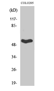
- Western Blot analysis of various cells using Septin 1 Polyclonal Antibody

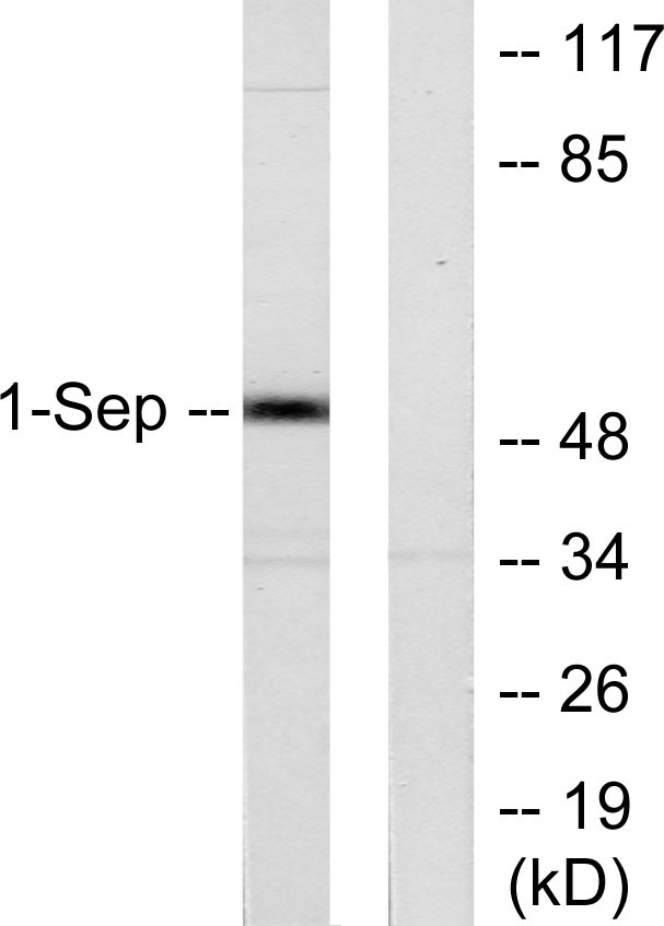
- Western blot analysis of lysates from Jurkat cells, using SEPT1 Antibody. The lane on the right is blocked with the synthesized peptide.

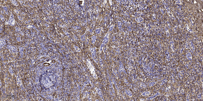
- Immunohistochemical analysis of paraffin-embedded human spleen tissue. 1,primary Antibody was diluted at 1:200(4° overnight). 2, Sodium citrate pH 6.0 was used for antigen retrieval(>98°C,20min). 3,Secondary antibody was diluted at 1:200


