TFDP1 Polyclonal Antibody
- Catalog No.:YT4611
- Applications:WB;IHC;IF;ELISA
- Reactivity:Human;Mouse
- Target:
- TFDP1
- Fields:
- >>Cell cycle;>>TGF-beta signaling pathway
- Gene Name:
- TFDP1
- Protein Name:
- Transcription factor Dp-1
- Human Gene Id:
- 7027
- Human Swiss Prot No:
- Q14186
- Mouse Swiss Prot No:
- Q08639
- Immunogen:
- The antiserum was produced against synthesized peptide derived from human DP-1. AA range:361-410
- Specificity:
- TFDP1 Polyclonal Antibody detects endogenous levels of TFDP1 protein.
- Formulation:
- Liquid in PBS containing 50% glycerol, 0.5% BSA and 0.02% sodium azide.
- Source:
- Polyclonal, Rabbit,IgG
- Dilution:
- WB 1:500 - 1:2000. IHC 1:100 - 1:300. IF 1:200 - 1:1000. ELISA: 1:20000. Not yet tested in other applications.
- Purification:
- The antibody was affinity-purified from rabbit antiserum by affinity-chromatography using epitope-specific immunogen.
- Concentration:
- 1 mg/ml
- Storage Stability:
- -15°C to -25°C/1 year(Do not lower than -25°C)
- Other Name:
- TFDP1;DP1;Transcription factor Dp-1;DRTF1-polypeptide 1;DRTF1;E2F dimerization partner 1
- Observed Band(KD):
- 55kD
- Background:
- This gene encodes a member of a family of transcription factors that heterodimerize with E2F proteins to enhance their DNA-binding activity and promote transcription from E2F target genes. The encoded protein functions as part of this complex to control the transcriptional activity of numerous genes involved in cell cycle progression from G1 to S phase. Alternative splicing results in multiple transcript variants. Pseudogenes of this gene are found on chromosomes 1, 15, and X.[provided by RefSeq, Jan 2009],
- Function:
- function:Can stimulate E2F-dependent transcription. Binds DNA cooperatively with E2F family members through the E2 recognition site, 5'-TTTC[CG]CGC-3', found in the promoter region of a number of genes whose products are involved in cell cycle regulation or in DNA replication. The DP2/E2F complex functions in the control of cell-cycle progression from G1 to S phase. The E2F-1/DP complex appears to mediate both cell proliferation and apoptosis.,induction:Down-regulated during differentiation.,miscellaneous:E2F/DP transactivation can be mediated by several cofactors including TBP, TFIIH, MDM2 and CBP.,PTM:Phosphorylation by E2F-1-bound cyclin A-CDK2, in the S phase, inhibits E2F-mediated DNA binding and transactivation.,similarity:Belongs to the E2F/DP family.,subunit:Component of the E2F/DP transcription factor complex. Forms heterodimers with E2F family members. The complex can interact
- Subcellular Location:
- Nucleus . Cytoplasm . Shuttles between the cytoplasm and nucleus and translocates into the nuclear compartment upon heterodimerization with E2F1. .
- Expression:
- Highest levels in muscle. Also expressed in brain, placenta, liver and kidney. Lower levels in lung and pancreas. Not detected in heart.
- June 19-2018
- WESTERN IMMUNOBLOTTING PROTOCOL
- June 19-2018
- IMMUNOHISTOCHEMISTRY-PARAFFIN PROTOCOL
- June 19-2018
- IMMUNOFLUORESCENCE PROTOCOL
- September 08-2020
- FLOW-CYTOMEYRT-PROTOCOL
- May 20-2022
- Cell-Based ELISA│解您多样本WB检测之困扰
- July 13-2018
- CELL-BASED-ELISA-PROTOCOL-FOR-ACETYL-PROTEIN
- July 13-2018
- CELL-BASED-ELISA-PROTOCOL-FOR-PHOSPHO-PROTEIN
- July 13-2018
- Antibody-FAQs
- Products Images
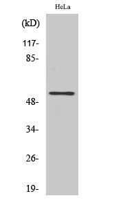
- Western Blot analysis of various cells using TFDP1 Polyclonal Antibody cells nucleus extracted by Minute TM Cytoplasmic and Nuclear Fractionation kit (SC-003,Inventbiotech,MN,USA).
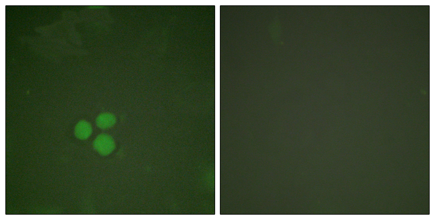
- Immunofluorescence analysis of HeLa cells, using DP-1 Antibody. The picture on the right is blocked with the synthesized peptide.
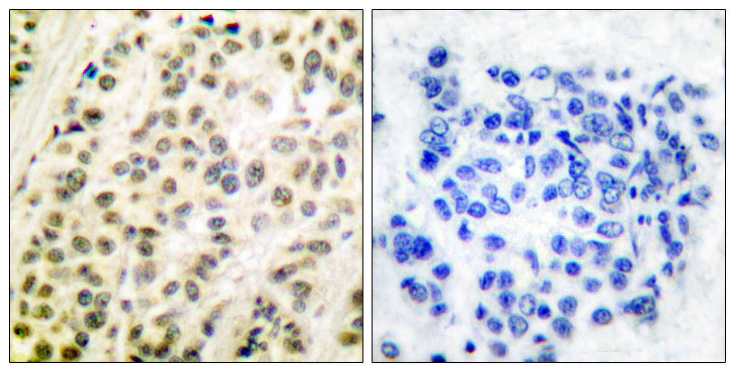
- Immunohistochemistry analysis of paraffin-embedded human breast carcinoma tissue, using DP-1 Antibody. The picture on the right is blocked with the synthesized peptide.
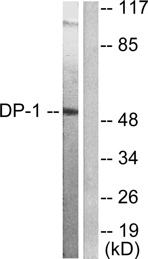
- Western blot analysis of lysates from HeLa cells, using DP-1 Antibody. The lane on the right is blocked with the synthesized peptide.
