Cleaved-Caspase-8 (D384) Polyclonal Antibody
- Catalog No.:YC0011
- Applications:WB;IHC;IF;ELISA
- Reactivity:Human;Rat;Mouse;
- Target:
- Caspase-8
- Fields:
- >>Platinum drug resistance;>>p53 signaling pathway;>>Apoptosis;>>Apoptosis - multiple species;>>Necroptosis;>>Toll-like receptor signaling pathway;>>NOD-like receptor signaling pathway;>>RIG-I-like receptor signaling pathway;>>C-type lectin receptor signaling pathway;>>IL-17 signaling pathway;>>TNF signaling pathway;>>Non-alcoholic fatty liver disease;>>Alcoholic liver disease;>>Alzheimer disease;>>Huntington disease;>>Pathways of neurodegeneration - multiple diseases;>>Pathogenic Escherichia coli infection;>>Salmonella infection;>>Legionellosis;>>Chagas disease;>>Toxoplasmosis;>>Tuberculosis;>>Hepatitis C;>>Hepatitis B;>>Measles;>>Human cytomegalovirus infection;>>Influenza A;>>Human papillomavirus infection;>>Kaposi sarcoma-associated herpesvirus infection;>>Herpes simplex virus 1 infection;>>Epstein-Barr virus infection;>>Human immunodeficiency virus 1 infection;>>Pathways in cancer;>>Viral carcinogenesis;>>Viral myocarditis;>>Lipid and atherosclerosis
- Gene Name:
- CASP8
- Protein Name:
- Caspase8
- Human Gene Id:
- 841
- Human Swiss Prot No:
- Q14790
- Mouse Swiss Prot No:
- O89110
- Immunogen:
- The antiserum was produced against synthesized peptide derived from human Caspase 8. AA range:335-384
- Specificity:
- Cleaved-Caspase-8 (D384) Polyclonal Antibody detects endogenous levels of fragment of activated Caspase-8 protein resulting from cleavage adjacent to D384.
- Formulation:
- Liquid in PBS containing 50% glycerol, 0.5% BSA and 0.02% sodium azide.
- Source:
- Polyclonal, Rabbit,IgG
- Dilution:
- WB 1:500-2000, IF 1:50-300, IHC 1:50-300
- Purification:
- The antibody was affinity-purified from rabbit antiserum by affinity-chromatography using epitope-specific immunogen.
- Concentration:
- 1 mg/ml
- Storage Stability:
- -15°C to -25°C/1 year(Do not lower than -25°C)
- Other Name:
- CASP8;MCH5;Caspase-8;CASP-8;Apoptotic cysteine protease;Apoptotic protease Mch-5;CAP4;FADD-homologous ICE/ced-3-like protease;FADD-like ICE;FLICE;ICE-like apoptotic protease 5;MORT1-associated ced-3 homolog;MACH
- Observed Band(KD):
- 47kD,55kD
- Background:
- This gene encodes a member of the cysteine-aspartic acid protease (caspase) family. Sequential activation of caspases plays a central role in the execution-phase of cell apoptosis. Caspases exist as inactive proenzymes composed of a prodomain, a large protease subunit, and a small protease subunit. Activation of caspases requires proteolytic processing at conserved internal aspartic residues to generate a heterodimeric enzyme consisting of the large and small subunits. This protein is involved in the programmed cell death induced by Fas and various apoptotic stimuli. The N-terminal FADD-like death effector domain of this protein suggests that it may interact with Fas-interacting protein FADD. This protein was detected in the insoluble fraction of the affected brain region from Huntington disease patients but not in those from normal controls, which implicated the role in neurodegenerative diseases. Many alt
- Function:
- catalytic activity:Strict requirement for Asp at position P1 and has a preferred cleavage sequence of (Leu/Asp/Val)-Glu-Thr-Asp-|-(Gly/Ser/Ala).,disease:Defects in CASP8 are the cause of caspase-8 deficiency (CASP8D) [MIM:607271]. CASP8D is a disorder resembling autoimmune lymphoproliferative syndrome (ALPS). It is characterized by lymphadenopathy, splenomegaly, and defective CD95-induced apoptosis of peripheral blood lymphocytes (PBLs). It leads to defects in activation of T-lymphocytes, B-lymphocytes, and natural killer cells leading to immunodeficiency characterized by recurrent sinopulmonary and herpes simplex virus infections and poor responses to immunization.,domain:Isoform 9 contains a N-terminal extension that is required for interaction with the BCAP31 complex.,function:Most upstream protease of the activation cascade of caspases responsible for the TNFRSF6/FAS mediated and TNF
- Subcellular Location:
- Cytoplasm . Nucleus .
- Expression:
- Isoform 1, isoform 5 and isoform 7 are expressed in a wide variety of tissues. Highest expression in peripheral blood leukocytes, spleen, thymus and liver. Barely detectable in brain, testis and skeletal muscle.
Terbinafine prevents colorectal cancer growth by inducing dNTP starvation and reducing immune suppression WB Human 1:1000 /HCT116, HT-29 cells
Ganoderic Acids Prevent Renal Ischemia Reperfusion Injury by Inhibiting Inflammation and Apoptosis. INTERNATIONAL JOURNAL OF MOLECULAR SCIENCES Int J Mol Sci. 2021 Jan;22(19):10229 WB Rat Renal tissues NRK-52E cell
miR-378 inhibits cell growth and enhances apoptosis in human myelodysplastic syndromes. INTERNATIONAL JOURNAL OF ONCOLOGY 2016 Nov 01 WB Human SKM-1 cell
Targeting FoxM1 by thiostrepton inhibits growth and induces apoptosis of laryngeal squamous cell carcinoma. JOURNAL OF CANCER RESEARCH AND CLINICAL ONCOLOGY 2014 Nov 13 WB Human Hep-2 cell
Protective effect of luteolin on cigarette smoke extract‑induced cellular toxicity and apoptosis in normal human bronchial epithelial cells via the Nrf2 pathway. ONCOLOGY REPORTS Oncol Rep. 2014 Apr;31(4):1855-1862 WB Human NHBE cell
Coptisine Induces Apoptosis in Human Hepatoma Cells Through Activating 67-kDa Laminin Receptor/cGMP Signaling. Frontiers in Pharmacology 2018 May 18 WB,IHC Human 1:500,1:500 SMMC7721 cell, HepG2 Cell
Down-regulation of FoxM1 by thiostrepton or small interfering RNA inhibits proliferation, transformation ability and angiogenesis, and induces apoptosis of nasopharyngeal carcinoma cells. International Journal of Clinical and Experimental Pathology Int J Clin Exp Patho. 2014; 7(9): 5450–5460 WB Human Nasopharyngeal carcinoma (NPC) cell
Synergistic antitumor effect of suberoylanilide hydroxamic acid and cisplatin in osteosarcoma cells. Oncology Letters 2018 Jul 27 WB Human 143B cell
Coptisine Induces Apoptosis in Human Hepatoma Cells Through Activating 67-kDa Laminin Receptor/cGMP Signaling. Frontiers in Pharmacology 2018 May 18 WB Human 1:500 SMMC7721 cell, HepG2 Cell
A novel monoclonal antibody associated with glucoside kills gastric adenocarcinoma AGS cells based on glycosylation target JOURNAL OF CELLULAR AND MOLECULAR MEDICINE Heng Xu WB Human
The outer membrane protein Tp92 of Treponema pallidum delays human neutrophil apoptosis via the ERK, PI3K/Akt, and NF-κB pathways. MOLECULAR MICROBIOLOGY Yimou Wu WB Human human polymorphonuclear neutrophils (hPMNs)
hPMSCs Regulate the Level of TNF-α and IL-10 in Th1 Cells and Improve Hepatic Injury in a GVHD Mouse Model via CD73/ADO/Fyn/Nrf2 Axis. INFLAMMATION Luan Xiying IF Human human placental-derived MSCs (hPMSCs)
- June 19-2018
- WESTERN IMMUNOBLOTTING PROTOCOL
- June 19-2018
- IMMUNOHISTOCHEMISTRY-PARAFFIN PROTOCOL
- June 19-2018
- IMMUNOFLUORESCENCE PROTOCOL
- September 08-2020
- FLOW-CYTOMEYRT-PROTOCOL
- May 20-2022
- Cell-Based ELISA│解您多样本WB检测之困扰
- July 13-2018
- CELL-BASED-ELISA-PROTOCOL-FOR-ACETYL-PROTEIN
- July 13-2018
- CELL-BASED-ELISA-PROTOCOL-FOR-PHOSPHO-PROTEIN
- July 13-2018
- Antibody-FAQs
- Products Images
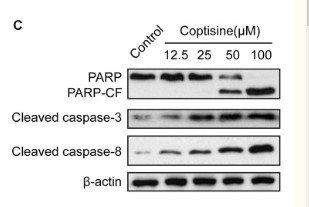
- Zhou, Li, et al. "Coptisine induces apoptosis in human hepatoma cells through activating 67-kDa laminin receptor/cGMP signaling." Frontiers in pharmacology 9 (2018).

- Hou, Mengyi, et al. "Synergistic antitumor effect of suberoylanilide hydroxamic acid and cisplatin in osteosarcoma cells." Oncology letters 16.4 (2018): 4663-4670.
poly-ihc-human-liver-cancer.jpg)
- Immunohistochemical analysis of paraffin-embedded Human-liver-cancer tissue. 1,Cleaved-Caspase-8 (D384) Polyclonal Antibody was diluted at 1:200(4°C,overnight). 2, Sodium citrate pH 6.0 was used for antibody retrieval(>98°C,20min). 3,Secondary antibody was diluted at 1:200(room tempeRature, 30min). Negative control was used by secondary antibody only.
poly-ihc-human-kidney.jpg)
- Immunohistochemical analysis of paraffin-embedded Human-kidney tissue. 1,Cleaved-Caspase-8 (D384) Polyclonal Antibody was diluted at 1:200(4°C,overnight). 2, Sodium citrate pH 6.0 was used for antibody retrieval(>98°C,20min). 3,Secondary antibody was diluted at 1:200(room tempeRature, 30min). Negative control was used by secondary antibody only.
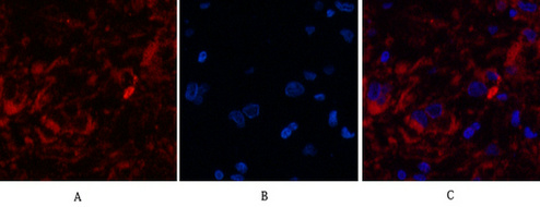
- Immunofluorescence analysis of Human-breast-cancer tissue. 1,Cleaved-Caspase-8 (D384) Polyclonal Antibody(red) was diluted at 1:200(4°C,overnight). 2, Cy3 labled Secondary antibody was diluted at 1:300(room temperature, 50min).3, Picture B: DAPI(blue) 10min. Picture A:Target. Picture B: DAPI. Picture C: merge of A+B
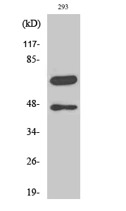
- Western Blot analysis of various cells using Cleaved-Caspase-8 (D384) Polyclonal Antibody
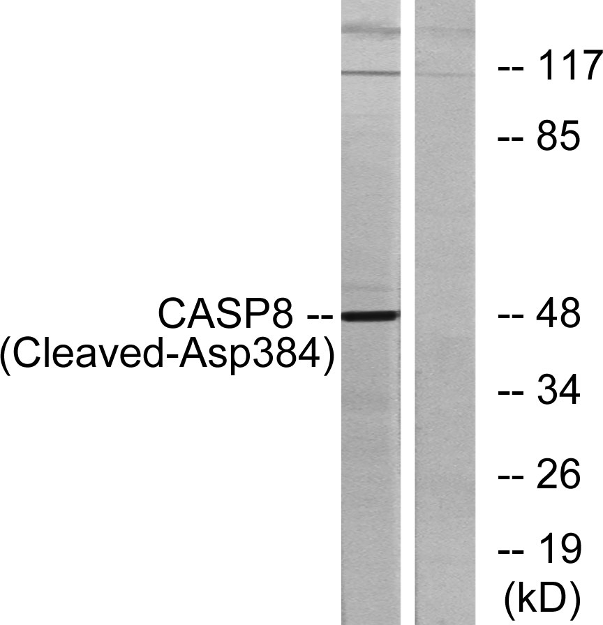
- Western blot analysis of lysates from 293 cells, treated with etoposide 25uM 1h, using Caspase 8 (Cleaved-Asp384) Antibody. The lane on the right is blocked with the synthesized peptide.


