E2F-1 (Acetyl-K125) Polyclonal Antibody
- Catalog No.:YK0088
- Applications:WB;ELISA
- Reactivity:Human:K125;Mouse:K120;Rat:K123
- Target:
- E2F-1
- Fields:
- >>Endocrine resistance;>>Cell cycle;>>Mitophagy - animal;>>Cellular senescence;>>Cushing syndrome;>>Hepatitis C;>>Hepatitis B;>>Human cytomegalovirus infection;>>Human papillomavirus infection;>>Human T-cell leukemia virus 1 infection;>>Kaposi sarcoma-associated herpesvirus infection;>>Epstein-Barr virus infection;>>Pathways in cancer;>>MicroRNAs in cancer;>>Chemical carcinogenesis - receptor activation;>>Pancreatic cancer;>>Glioma;>>Prostate cancer;>>Melanoma;>>Bladder cancer;>>Chronic myeloid leukemia;>>Small cell lung cancer;>>Non-small cell lung cancer;>>Breast cancer;>>Hepatocellular carcinoma;>>Gastric cancer
- Gene Name:
- E2F1 RBBP3
- Protein Name:
- E2F-1
- Human Gene Id:
- 1869
- Human Swiss Prot No:
- Q01094
- Mouse Swiss Prot No:
- Q61501
- Immunogen:
- Synthesized Acetyl peptide derived from human E2F-1. at AA range: K125
- Specificity:
- This antibody detects endogenous levels of E2F-1 at Human:K125;Mouse:K120;Rat:K123, It doesn't reacte with total protein.
- Formulation:
- Liquid in PBS containing 50% glycerol, 0.5% BSA and 0.02% sodium azide.
- Source:
- Polyclonal, Rabbit,IgG
- Dilution:
- WB 1:500 - 1:2000. ELISA: 1:20000. Not yet tested in other applications.
- Purification:
- The antibody was affinity-purified from rabbit antiserum by affinity-chromatography using epitope-specific immunogen.
- Concentration:
- 1 mg/ml
- Storage Stability:
- -15°C to -25°C/1 year(Do not lower than -25°C)
- Other Name:
- Transcription factor E2F1 (E2F-1) (PBR3) (Retinoblastoma-associated protein 1) (RBAP-1) (Retinoblastoma-binding protein 3) (RBBP-3) (pRB-binding protein E2F-1)
- Observed Band(KD):
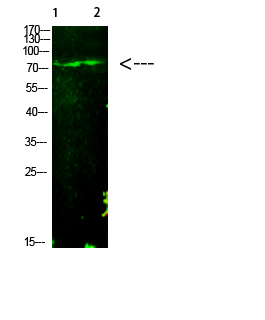
- 60kD
- Background:
- The protein encoded by this gene is a member of the E2F family of transcription factors. The E2F family plays a crucial role in the control of cell cycle and action of tumor suppressor proteins and is also a target of the transforming proteins of small DNA tumor viruses. The E2F proteins contain several evolutionally conserved domains found in most members of the family. These domains include a DNA binding domain, a dimerization domain which determines interaction with the differentiation regulated transcription factor proteins (DP), a transactivation domain enriched in acidic amino acids, and a tumor suppressor protein association domain which is embedded within the transactivation domain. This protein and another 2 members, E2F2 and E2F3, have an additional cyclin binding domain. This protein binds preferentially to retinoblastoma protein pRB in a cell-cycle dependent manner. It can media
- Function:
- function:Transcription activator that binds DNA cooperatively with dp proteins through the E2 recognition site, 5'-TTTC[CG]CGC-3' found in the promoter region of a number of genes whose products are involved in cell cycle regulation or in DNA replication. The DRTF1/E2F complex functions in the control of cell-cycle progression from G1 to S phase. E2F-1 binds preferentially RB1 protein, in a cell-cycle dependent manner. It can mediate both cell proliferation and p53-dependent apoptosis.,PTM:Phosphorylated by CDK2 and cyclin A-CDK2 in the S-phase.,similarity:Belongs to the E2F/DP family.,subunit:Component of the DRTF1/E2F transcription factor complex. Forms heterodimers with DP family members. The E2F-1 complex binds specifically hypophosphorylated retinoblastoma protein RB1. During the cell cycle, RB1 becomes phosphorylated in mid-to-late G1 phase, detaches from the DRTF1/E2F complex, ren
- Subcellular Location:
- Nucleus .
- Expression:
- Brain,Epithelium,Pancreas,Skin,
- June 19-2018
- WESTERN IMMUNOBLOTTING PROTOCOL
- June 19-2018
- IMMUNOHISTOCHEMISTRY-PARAFFIN PROTOCOL
- June 19-2018
- IMMUNOFLUORESCENCE PROTOCOL
- September 08-2020
- FLOW-CYTOMEYRT-PROTOCOL
- May 20-2022
- Cell-Based ELISA│解您多样本WB检测之困扰
- July 13-2018
- CELL-BASED-ELISA-PROTOCOL-FOR-ACETYL-PROTEIN
- July 13-2018
- CELL-BASED-ELISA-PROTOCOL-FOR-PHOSPHO-PROTEIN
- July 13-2018
- Antibody-FAQs
- Products Images

- Western Blot analysis of 1,mouse-liver 2,hela cells using primary antibody diluted at 1:1000(4°C overnight). Secondary antibody:Goat Anti-rabbit IgG IRDye 800( diluted at 1:5000, 25°C, 1 hour)


