CD4 Monoclonal Antibody
- Catalog No.:YM0124
- Applications:FCM;ELISA
- Reactivity:Human
- Target:
- CD4
- Fields:
- >>Viral life cycle - HIV-1;>>Cytokine-cytokine receptor interaction;>>Cell adhesion molecules;>>Antigen processing and presentation;>>Hematopoietic cell lineage;>>Th1 and Th2 cell differentiation;>>Th17 cell differentiation;>>T cell receptor signaling pathway;>>Yersinia infection;>>Human T-cell leukemia virus 1 infection;>>Human immunodeficiency virus 1 infection;>>PD-L1 expression and PD-1 checkpoint pathway in cancer;>>Primary immunodeficiency
- Gene Name:
- CD4
- Protein Name:
- T-cell surface glycoprotein CD4
- Human Gene Id:
- 920
- Human Swiss Prot No:
- P01730
- Mouse Swiss Prot No:
- P06332
- Immunogen:
- Purified recombinant fragment of human CD4 expressed in E. Coli.
- Specificity:
- CD4 Monoclonal Antibody detects endogenous levels of CD4 protein.
- Formulation:
- Liquid in PBS containing 50% glycerol, 0.5% BSA and 0.02% sodium azide.
- Source:
- Monoclonal, Mouse
- Dilution:
- Flow cytometry: 1:200 - 1:400. ELISA: 1:10000. Not yet tested in other applications.
- Purification:
- Affinity purification
- Concentration:
- 0.5 mg/ml
- Storage Stability:
- -15°C to -25°C/1 year(Do not lower than -25°C)
- Other Name:
- CD4;T-cell surface glycoprotein CD4;T-cell surface antigen T4/Leu-3;CD antigen CD4
- References:
- 1. M Benkirane J Virol. 1995 November; 69(11): 6898–6903.
- Background:
- This gene encodes a membrane glycoprotein of T lymphocytes that interacts with major histocompatibility complex class II antigenes and is also a receptor for the human immunodeficiency virus. This gene is expressed not only in T lymphocytes, but also in B cells, macrophages, and granulocytes. It is also expressed in specific regions of the brain. The protein functions to initiate or augment the early phase of T-cell activation, and may function as an important mediator of indirect neuronal damage in infectious and immune-mediated diseases of the central nervous system. Multiple alternatively spliced transcript variants encoding different isoforms have been identified in this gene. [provided by RefSeq, Aug 2010],
- Function:
- function:Accessory protein for MHC class-II antigen/T-cell receptor interaction. May regulate T-cell activation. Induces the aggregation of lipid rafts.,miscellaneous:Primary receptor for HIV-1.,online information:CD4 entry,PTM:Palmitoylation and association with LCK contribute to the enrichment of CD4 in lipid rafts.,similarity:Contains 1 Ig-like V-type (immunoglobulin-like) domain.,similarity:Contains 3 Ig-like C2-type (immunoglobulin-like) domains.,subcellular location:Localizes to lipid rafts. Removed from plasma membrane by HIV-1 Nef protein that increases clathrin-dependent endocytosis of this antigen to target it to lysosomal degradation. Cell surface expression is also down-modulated by HIV-1 Envelope polyprotein gp160 that interacts with, and sequesters CD4 in the endoplasmic reticulum.,subunit:Associates with LCK. Binds to HIV-1 gp120 and to P4HB/PDI and upon HIV-1 binding to t
- Subcellular Location:
- Cell membrane ; Single-pass type I membrane protein . Localizes to lipid rafts (PubMed:12517957, PubMed:9168119). Removed from plasma membrane by HIV-1 Nef protein that increases clathrin-dependent endocytosis of this antigen to target it to lysosomal degradation. Cell surface expression is also down-modulated by HIV-1 Envelope polyprotein gp160 that interacts with, and sequesters CD4 in the endoplasmic reticulum.
- Expression:
- Highly expressed in T-helper cells. The presence of CD4 is a hallmark of T-helper cells which are specialized in the activation and growth of cytotoxic T-cells, regulation of B cells, or activation of phagocytes. CD4 is also present in other immune cells such as macrophages, dendritic cells or NK cells.
Effect of montelukast combined with methylprednisolone for the treatment of mycoplasma pneumonia:. JOURNAL OF INTERNATIONAL MEDICAL RESEARCH 2019 May 09 FC Human T cell
- June 19-2018
- WESTERN IMMUNOBLOTTING PROTOCOL
- June 19-2018
- IMMUNOHISTOCHEMISTRY-PARAFFIN PROTOCOL
- June 19-2018
- IMMUNOFLUORESCENCE PROTOCOL
- September 08-2020
- FLOW-CYTOMEYRT-PROTOCOL
- May 20-2022
- Cell-Based ELISA│解您多样本WB检测之困扰
- July 13-2018
- CELL-BASED-ELISA-PROTOCOL-FOR-ACETYL-PROTEIN
- July 13-2018
- CELL-BASED-ELISA-PROTOCOL-FOR-PHOSPHO-PROTEIN
- July 13-2018
- Antibody-FAQs
- Products Images
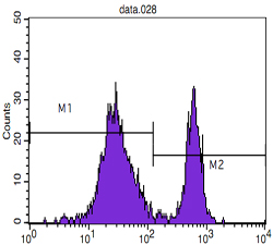
- Flow cytometric analysis of blood T cells using CD4 Monoclonal Antibody (M2) and negative control (M1).


