eIF4E Monoclonal Antibody
- Catalog No.:YM0214
- Applications:WB;IHC;IF;FCM;ELISA
- Reactivity:Human
- Target:
- eIF4E
- Fields:
- >>EGFR tyrosine kinase inhibitor resistance;>>HIF-1 signaling pathway;>>mTOR signaling pathway;>>PI3K-Akt signaling pathway;>>Longevity regulating pathway;>>Insulin signaling pathway
- Gene Name:
- EIF4E
- Protein Name:
- Eukaryotic translation initiation factor 4E
- Human Gene Id:
- 1977
- Human Swiss Prot No:
- P06730
- Mouse Swiss Prot No:
- P63073
- Immunogen:
- Purified recombinant fragment of human eIF4E expressed in E. Coli.
- Specificity:
- eIF4E Monoclonal Antibody detects endogenous levels of eIF4E protein.
- Formulation:
- Liquid in PBS containing 50% glycerol, 0.5% BSA and 0.02% sodium azide.
- Source:
- Monoclonal, Mouse
- Dilution:
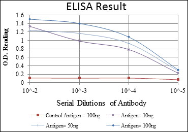
- WB 1:500 - 1:2000. IHC 1:200 - 1:1000. IF 1:200 - 1:1000. Flow cytometry: 1:200 - 1:400. ELISA: 1:10000. Not yet tested in other applications.
- Purification:
- Affinity purification
- Storage Stability:
- -15°C to -25°C/1 year(Do not lower than -25°C)
- Other Name:
- EIF4E;EIF4EL1;EIF4F;Eukaryotic translation initiation factor 4E;eIF-4E;eIF4E;eIF-4F 25 kDa subunit;mRNA cap-binding protein
- Molecular Weight(Da):
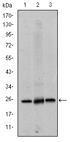
- 25kD
- References:
- 1. Ann Surg Oncol. 2008 Nov;15(11):3207-15.
2. J Biol Chem. 2008 Sep 12;283(37):25227-37.
- Background:
- The protein encoded by this gene is a component of the eukaryotic translation initiation factor 4F complex, which recognizes the 7-methylguanosine cap structure at the 5' end of messenger RNAs. The encoded protein aids in translation initiation by recruiting ribosomes to the 5'-cap structure. Association of this protein with the 4F complex is the rate-limiting step in translation initiation. This gene acts as a proto-oncogene, and its expression and activation is associated with transformation and tumorigenesis. Several pseudogenes of this gene are found on other chromosomes. Alternative splicing results in multiple transcript variants. [provided by RefSeq, Sep 2015],
- Function:
- caution:Was originally thought to be phosphorylated on Ser-53 (PubMed:3112145); this was later shown to be wrong (PubMed:7665584).,function:Recognizes and binds the 7-methylguanosine-containing mRNA cap during an early step in the initiation of protein synthesis and facilitates ribosome binding by inducing the unwinding of the mRNAs secondary structures.,PTM:Phosphorylation increases the ability of the protein to bind to mRNA caps and to form the eIF4F complex.,similarity:Belongs to the eukaryotic initiation factor 4E family.,subunit:eIF4F is a multi-subunit complex, the composition of which varies with external and internal environmental conditions. It is composed of at least EIF4A, EIF4E and EIF4G1/EIF4G3. EIF4E is also known to interact with other partners. The interaction with EIF4ENIF1 mediates the import into the nucleus. Nonphosphorylated EIF4EBP1, EIF4EBP2 and EIF4EBP3 compete wi
- Subcellular Location:
- Cytoplasm, P-body . Cytoplasm . Cytoplasm, Stress granule . Nucleus . Interaction with EIF4ENIF1/4E-T is required for localization to processing bodies (P-bodies) (PubMed:16157702, PubMed:24335285, PubMed:25923732). Imported in the nucleus via interaction with EIF4ENIF1/4E-T via a piggy-back mechanism (PubMed:10856257). .
- Expression:
- Brain,Fetal brain,Placenta,Pooled,Small intestine,Testis,
- June 19-2018
- WESTERN IMMUNOBLOTTING PROTOCOL
- June 19-2018
- IMMUNOHISTOCHEMISTRY-PARAFFIN PROTOCOL
- June 19-2018
- IMMUNOFLUORESCENCE PROTOCOL
- September 08-2020
- FLOW-CYTOMEYRT-PROTOCOL
- May 20-2022
- Cell-Based ELISA│解您多样本WB检测之困扰
- July 13-2018
- CELL-BASED-ELISA-PROTOCOL-FOR-ACETYL-PROTEIN
- July 13-2018
- CELL-BASED-ELISA-PROTOCOL-FOR-PHOSPHO-PROTEIN
- July 13-2018
- Antibody-FAQs
- Products Images

- Western Blot analysis using eIF4E Monoclonal Antibody against HeLa (1), HEK293 (2) and K562 (3) cell lysate.

- Immunohistochemistry analysis of paraffin-embedded liver cancer (left) and submaxillary tumor (right) with DAB staining using eIF4E Monoclonal Antibody.

- Immunofluorescence analysis of GC-7901 cells using eIF4E Monoclonal Antibody (green). Blue: DRAQ5 fluorescent DNA dye. Red: Actin filaments have been labeled with Alexa Fluor-555 phalloidin.

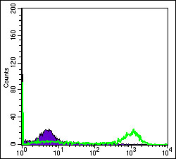
- Flow cytometric analysis of Hela cells using eIF4E Monoclonal Antibody (green) and negative control (purple).


