HSP27 Monoclonal Antibody
- Catalog No.:YM0339
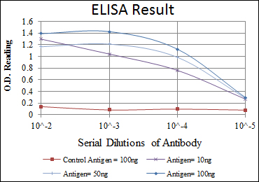
- Applications:WB;IHC;IF;FCM;ELISA
- Reactivity:Human;Rat
- Target:
- HSP27
- Fields:
- >>MAPK signaling pathway;>>VEGF signaling pathway;>>Amoebiasis
- Gene Name:
- HSPB1
- Protein Name:
- Heat shock protein beta-1
- Human Gene Id:
- 3315
- Human Swiss Prot No:
- P04792
- Mouse Swiss Prot No:
- P14602
- Rat Gene Id:
- 24471
- Rat Swiss Prot No:
- P42930
- Immunogen:
- Purified recombinant fragment of human HSP27 expressed in E. Coli.
- Specificity:
- HSP27 Monoclonal Antibody detects endogenous levels of HSP27 protein.
- Formulation:
- Liquid in PBS containing 50% glycerol, 0.5% BSA and 0.02% sodium azide.
- Source:
- Monoclonal, Mouse
- Dilution:
- WB 1:500 - 1:2000. IHC 1:200 - 1:1000. IF 1:200 - 1:1000. Flow cytometry: 1:200 - 1:400. ELISA: 1:10000. Not yet tested in other applications.
- Purification:
- Affinity purification
- Storage Stability:
- -15°C to -25°C/1 year(Do not lower than -25°C)
- Other Name:
- HSPB1;HSP27;HSP28;Heat shock protein beta-1;HspB1;28 kDa heat shock protein;Estrogen-regulated 24 kDa protein;Heat shock 27 kDa protein;HSP 27;Stress-responsive protein 27;SRP27
- Molecular Weight(Da):
- 23kD
- References:
- 1. Clin Cancer Res. 2008 Dec 15;14(24):8279-87.
2. Cell Signal. 2009 Jan;21(1):143-50.
- Background:
- The protein encoded by this gene is induced by environmental stress and developmental changes. The encoded protein is involved in stress resistance and actin organization and translocates from the cytoplasm to the nucleus upon stress induction. Defects in this gene are a cause of Charcot-Marie-Tooth disease type 2F (CMT2F) and distal hereditary motor neuropathy (dHMN). [provided by RefSeq, Oct 2008],
- Function:
- disease:Defects in HSPB1 are a cause of distal hereditary motor neuronopathy type 2B (HMN2B) [MIM:608634]. Distal hereditary motor neuronopathies constitute a heterogeneous group of neuromuscular disorders caused by selective impairment of motor neurons in the anterior horn of the spinal cord, without sensory deficit in the posterior horn. The overall clinical picture consists of a classical distal muscular atrophy syndrome in the legs without clinical sensory loss. The disease starts with weakness and wasting of distal muscles of the anterior tibial and peroneal compartments of the legs. Later on, weakness and atrophy may expand to the proximal muscles of the lower limbs and/or to the distal upper limbs.,disease:Defects in HSPB1 are the cause of Charcot-Marie-Tooth disease type 2F (CMT2F) [MIM:606595]. CMT2F is a form of Charcot-Marie-Tooth disease, the most common inherited disorder of
- Subcellular Location:
- Cytoplasm . Nucleus . Cytoplasm, cytoskeleton, spindle . Cytoplasmic in interphase cells. Colocalizes with mitotic spindles in mitotic cells. Translocates to the nucleus during heat shock and resides in sub-nuclear structures known as SC35 speckles or nuclear splicing speckles. .
- Expression:
- Detected in all tissues tested: skeletal muscle, heart, aorta, large intestine, small intestine, stomach, esophagus, bladder, adrenal gland, thyroid, pancreas, testis, adipose tissue, kidney, liver, spleen, cerebral cortex, blood serum and cerebrospinal fluid. Highest levels are found in the heart and in tissues composed of striated and smooth muscle.
- June 19-2018
- WESTERN IMMUNOBLOTTING PROTOCOL
- June 19-2018
- IMMUNOHISTOCHEMISTRY-PARAFFIN PROTOCOL
- June 19-2018
- IMMUNOFLUORESCENCE PROTOCOL
- September 08-2020
- FLOW-CYTOMEYRT-PROTOCOL
- May 20-2022
- Cell-Based ELISA│解您多样本WB检测之困扰
- July 13-2018
- CELL-BASED-ELISA-PROTOCOL-FOR-ACETYL-PROTEIN
- July 13-2018
- CELL-BASED-ELISA-PROTOCOL-FOR-PHOSPHO-PROTEIN
- July 13-2018
- Antibody-FAQs
- Products Images
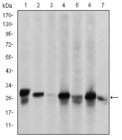
- Western Blot analysis using HSP27 Monoclonal Antibody against HeLa (1), A549 (2), Jurkat (3), A431 (4), HEK293(5), HepG2 (6) and PC-12 (7) cell lysate.
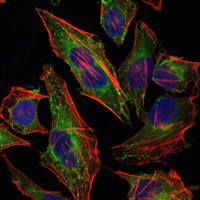
- Immunofluorescence analysis of Hela cells using HSP27 Monoclonal Antibody (green). Blue: DRAQ5 fluorescent DNA dye. Red: Actin filaments have been labeled with Alexa Fluor-555 phalloidin.
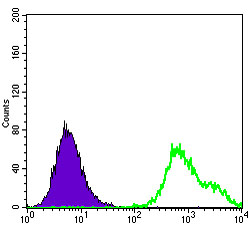
- Flow cytometric analysis of HepG2 cells using HSP27 Monoclonal Antibody (green) and negative control (purple).
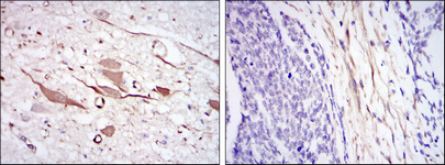
- Immunohistochemistry analysis of paraffin-embedded brain tissues (left) and esophageal cancer tissues (right) with DAB staining using HSP27 Monoclonal Antibody


