HXK I Monoclonal Antibody
- Catalog No.:YM0349
- Applications:WB;IHC;IF;FCM;ELISA
- Reactivity:Human;Mouse;Rat
- Target:
- HXK I
- Fields:
- >>Glycolysis / Gluconeogenesis;>>Fructose and mannose metabolism;>>Galactose metabolism;>>Starch and sucrose metabolism;>>Amino sugar and nucleotide sugar metabolism;>>Neomycin, kanamycin and gentamicin biosynthesis;>>Metabolic pathways;>>Carbon metabolism;>>Biosynthesis of nucleotide sugars;>>HIF-1 signaling pathway;>>Insulin signaling pathway;>>Type II diabetes mellitus;>>Carbohydrate digestion and absorption;>>Shigellosis;>>Central carbon metabolism in cancer
- Gene Name:
- HK1
- Protein Name:
- Hexokinase-1
- Human Gene Id:
- 3098
- Human Swiss Prot No:
- P19367
- Mouse Swiss Prot No:
- P17710
- Rat Gene Id:
- 25058
- Rat Swiss Prot No:
- P05708
- Immunogen:
- Purified recombinant fragment of human HXK I expressed in E. Coli.
- Specificity:
- HXK I Monoclonal Antibody detects endogenous levels of HXK I protein.
- Formulation:
- Liquid in PBS containing 50% glycerol, 0.5% BSA and 0.02% sodium azide.
- Source:
- Monoclonal, Mouse
- Dilution:
- WB 1:500 - 1:2000. IHC 1:200 - 1:1000. IF 1:200 - 1:1000. Flow cytometry: 1:200 - 1:400. ELISA: 1:10000. Not yet tested in other applications.
- Purification:
- Affinity purification
- Storage Stability:
- -15°C to -25°C/1 year(Do not lower than -25°C)
- Other Name:
- HK1;Hexokinase-1;Brain form hexokinase;Hexokinase type I;HK I
- Molecular Weight(Da):
- 102kD
- References:
- 1. Cell. 2005 Sep 23;122(6):957-68.
2. Am J Hum Genet. 2006 Jan;78(1):78-88.
- Background:
- Hexokinases phosphorylate glucose to produce glucose-6-phosphate, the first step in most glucose metabolism pathways. This gene encodes a ubiquitous form of hexokinase which localizes to the outer membrane of mitochondria. Mutations in this gene have been associated with hemolytic anemia due to hexokinase deficiency. Alternative splicing of this gene results in several transcript variants which encode different isoforms, some of which are tissue-specific. [provided by RefSeq, Apr 2016],
- Function:
- catalytic activity:ATP + D-hexose = ADP + D-hexose 6-phosphate.,disease:Defects in HK1 are the cause of hexokinase deficiency [MIM:235700]. Hexokinase deficiency is a rare autosomal recessive disease with nonspherocytic hemolytic anemia as the predominant clinical feature.,domain:The N- and C-terminal halves of this hexokinase show extensive sequence similarity to each other. The catalytic activity is associated with the C-terminus while regulatory function is associated with the N-terminus.,enzyme regulation:Hexokinase is an allosteric enzyme inhibited by its product Glc-6-P.,miscellaneous:In vertebrates there are four major glucose-phosphorylating isoenzymes, designated hexokinase I, II, III and IV (glucokinase).,online information:Hexokinase entry,pathway:Carbohydrate metabolism; hexose metabolism.,similarity:Belongs to the hexokinase family.,subcellular location:Its hydrophobic N-ter
- Subcellular Location:
- Mitochondrion outer membrane ; Peripheral membrane protein . Cytoplasm, cytosol . The mitochondrial-binding peptide (MBP) region promotes association with the mitochondrial outer membrane (Probable). Dissociates from the mitochondrial outer membrane following inhibition by N-acetyl-D-glucosamine, leading to relocation to the cytosol (PubMed:27374331). .
- Expression:
- Isoform 2: Erythrocyte specific (Ref.6). Isoform 3: Testis-specific (PubMed:10978502). Isoform 4: Testis-specific (PubMed:10978502).
- June 19-2018
- WESTERN IMMUNOBLOTTING PROTOCOL
- June 19-2018
- IMMUNOHISTOCHEMISTRY-PARAFFIN PROTOCOL
- June 19-2018
- IMMUNOFLUORESCENCE PROTOCOL
- September 08-2020
- FLOW-CYTOMEYRT-PROTOCOL
- May 20-2022
- Cell-Based ELISA│解您多样本WB检测之困扰
- July 13-2018
- CELL-BASED-ELISA-PROTOCOL-FOR-ACETYL-PROTEIN
- July 13-2018
- CELL-BASED-ELISA-PROTOCOL-FOR-PHOSPHO-PROTEIN
- July 13-2018
- Antibody-FAQs
- Products Images

- Western Blot analysis using HXK I Monoclonal Antibody against Jurkat (1), HeLa (2), HepG2 (3) and NIH/3T3 (4) cell lysate.

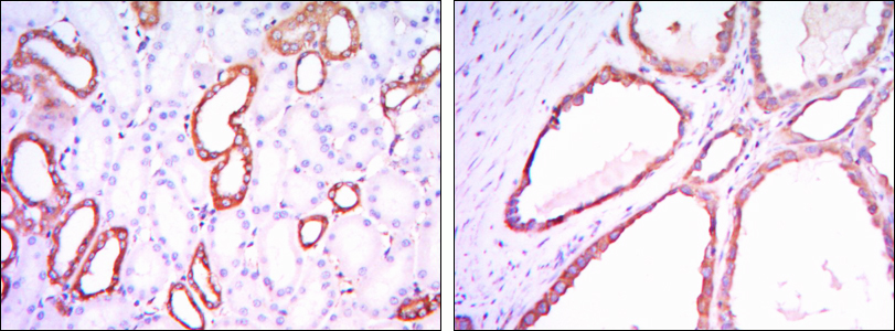
- Immunohistochemistry analysis of paraffin-embedded kidney tissues with DAB staining using HXK I Monoclonal Antibody.

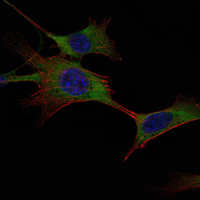
- Immunofluorescence analysis of NIH/3T3 cells using HXK I Monoclonal Antibody (green). Blue: DRAQ5 fluorescent DNA dye. Red: Actin filaments have been labeled with Alexa Fluor-555 phalloidin.

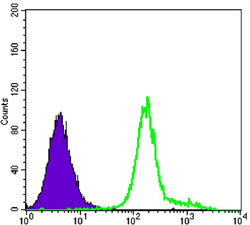
- Flow cytometric analysis of K562 cells using HXK I Monoclonal Antibody (green) and negative control (purple).


