OTX2 Monoclonal Antibody
- Catalog No.:YM0490
- Applications:WB;IHC;IF;FCM;ELISA
- Reactivity:Human
- Target:
- OTX2
- Gene Name:
- OTX2
- Protein Name:
- Homeobox protein OTX2
- Human Gene Id:
- 5015
- Human Swiss Prot No:
- P32243
- Mouse Swiss Prot No:
- P80206
- Immunogen:
- Purified recombinant fragment of human OTX2 expressed in E. Coli.
- Specificity:
- OTX2 Monoclonal Antibody detects endogenous levels of OTX2 protein.
- Formulation:
- Liquid in PBS containing 50% glycerol, 0.5% BSA and 0.02% sodium azide.
- Source:
- Monoclonal, Mouse
- Dilution:
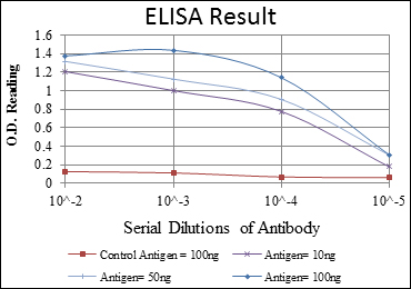
- WB 1:500 - 1:2000. IHC 1:200 - 1:1000. IF 1:200 - 1:1000. Flow cytometry: 1:200 - 1:400. ELISA: 1:10000. Not yet tested in other applications.
- Purification:
- Affinity purification
- Storage Stability:
- -15°C to -25°C/1 year(Do not lower than -25°C)
- Other Name:
- OTX2;Homeobox protein OTX2;Orthodenticle homolog 2
- Molecular Weight(Da):
- 32kD
- References:
- 1. Hum Mutat. 2008 Nov;29(11):E278-83.
2. Cancer Res. 2010 Jan 1;70(1):181-91.
- Background:
- This gene encodes a member of the bicoid subfamily of homeodomain-containing transcription factors. The encoded protein acts as a transcription factor and plays a role in brain, craniofacial, and sensory organ development. The encoded protein also influences the proliferation and differentiation of dopaminergic neuronal progenitor cells during mitosis. Mutations in this gene cause syndromic microphthalmia 5 (MCOPS5) and combined pituitary hormone deficiency 6 (CPHD6). This gene is also suspected of having an oncogenic role in medulloblastoma. Alternative splicing results in multiple transcript variants encoding distinct isoforms. Pseudogenes of this gene are known to exist on chromosomes two and nine. [provided by RefSeq, Jul 2012],
- Function:
- developmental stage:Embryo.,disease:Defects in OTX2 are the cause of microphthalmia syndromic type 5 (MCOPS5) [MIM:610125]. Microphthalmia is a clinically heterogeneous disorder of eye formation, ranging from small size of a single eye to complete bilateral absence of ocular tissues. Up to 80% of cases of microphthalia occur in association with syndromes that include non-ocular abnormalities. MCOPS5 patients manifest unilateral or bilateral microphthalmia/clinical anophthalmia and variable additional features including coloboma, microcornea, cataract, retinal dystrophy, hypoplasia or agenesis of the optic nerve, agenesis of the corpus callosum, developmental delay, joint laxity, hypotonia, and seizures.,function:Probably plays a role in the development of the brain and the sense organs. Can bind to the BCD target sequence (BTS): 5'-TCTAATCCC-3'.,similarity:Belongs to the paired homeobox
- Subcellular Location:
- Nucleus .
- Expression:
- Eye,Retina,
- June 19-2018
- WESTERN IMMUNOBLOTTING PROTOCOL
- June 19-2018
- IMMUNOHISTOCHEMISTRY-PARAFFIN PROTOCOL
- June 19-2018
- IMMUNOFLUORESCENCE PROTOCOL
- September 08-2020
- FLOW-CYTOMEYRT-PROTOCOL
- May 20-2022
- Cell-Based ELISA│解您多样本WB检测之困扰
- July 13-2018
- CELL-BASED-ELISA-PROTOCOL-FOR-ACETYL-PROTEIN
- July 13-2018
- CELL-BASED-ELISA-PROTOCOL-FOR-PHOSPHO-PROTEIN
- July 13-2018
- Antibody-FAQs
- Products Images
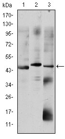
- Western Blot analysis using OTX2 Monoclonal Antibody against HepG2 (1), Jurkat (2), and NTERA-2 (3) cell lysate.
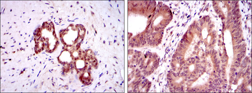
- Immunohistochemistry analysis of paraffin-embedded prostate tissues (left) and colon cancer tissues (right) with DAB staining using OTX2 Monoclonal Antibody.
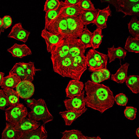
- Immunofluorescence analysis of HepG2 cells using OTX2 Monoclonal Antibody (green). Red: Actin filaments have been labeled with Alexa Fluor-555 phalloidin.
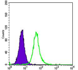
- Flow cytometric analysis of HepG2 cells using OTX2 Monoclonal Antibody (green) and negative control (purple).
