PEG10 Monoclonal Antibody
- Catalog No.:YM0515
- Applications:WB;IHC;IF;ELISA
- Reactivity:Human
- Target:
- PEG10
- Gene Name:
- PEG10
- Protein Name:
- Retrotransposon-derived protein PEG10
- Human Gene Id:
- 23089
- Human Swiss Prot No:
- Q86TG7
- Mouse Swiss Prot No:
- Q7TN75
- Immunogen:
- Purified recombinant fragment of PEG10 (aa1-120) expressed in E. Coli.
- Specificity:
- PEG10 Monoclonal Antibody detects endogenous levels of PEG10 protein.
- Formulation:
- Liquid in PBS containing 50% glycerol, 0.5% BSA and 0.02% sodium azide.
- Source:
- Monoclonal, Mouse
- Dilution:
- WB 1:500 - 1:2000. IHC 1:200 - 1:1000. IF 1:200 - 1:1000. ELISA: 1:10000. Not yet tested in other applications.
- Purification:
- Affinity purification
- Storage Stability:
- -15°C to -25°C/1 year(Do not lower than -25°C)
- Other Name:
- PEG10;EDR;KIAA1051;MAR2;MART2;MEF3L1;RGAG3;Retrotransposon-derived protein PEG10;Embryonal carcinoma differentiation-regulated protein;Mammalian retrotransposon-derived protein 2;Myelin expression factor 3-like protein 1;MEF3-lik
- Molecular Weight(Da):
- 80kD
- References:
- 1. Oncogene. 2007 Aug 23;26(39):5741-51.
2. FEBS Lett. 2008 Aug 6;582(18):2793-8.
- Background:
- This is a paternally expressed imprinted gene that is thought to have been derived from the Ty3/Gypsy family of retrotransposons. It contains two overlapping open reading frames, RF1 and RF2, and expresses two proteins: a shorter, gag-like protein (with a CCHC-type zinc finger domain) from RF1; and a longer, gag/pol-like fusion protein (with an additional aspartic protease motif) from RF1/RF2 by -1 translational frameshifting (-1 FS). While -1 FS has been observed in RNA viruses and transposons in both prokaryotes and eukaryotes, this gene represents the first example of -1 FS in a eukaryotic cellular gene. This gene is highly conserved across mammalian species and retains the heptanucleotide (GGGAAAC) and pseudoknot elements required for -1 FS. It is expressed in adult and embryonic tissues (most notably in placenta) and reported to have a role in cell proliferation, differentiation and apoptosis.
- Function:
- alternative products:The ribosomal frameshifting efficiency yield up to 66% of protein RF1/RF2 compared to RF1,developmental stage:Expressed in placenta during the first trimester of gestation (at protein level). In placenta, down-regulated at early hypoxic phase, and highly activated at 11-12 week of gestation.,function:Prevents apoptosis in hepatocellular carcinoma (HCC) cells through interaction with SIAH1, a mediator of apoptosis. May also have a role in cell growth promotion and hepatoma formation. Inhibits the TGF-beta signaling by interacting with the TGF-beta receptor ALK1. When overexpressed, induces the formation of cellular extension, such as filipodia in association with ALK1. Involved at the immediate early stage of adipocyte differentiation (By similarity). May bind to the 5'-GCCTGTCTTT-3' DNA sequence of the MB1 domain in the myelin basic protein (MBP) promoter.,induction:
- Subcellular Location:
- Extracellular vesicle membrane . Cytoplasm . Nucleus . Forms virion-like extracellular vesicles that are released from cells (PubMed:34413232). Detected predominantly in the cytoplasm of breast and prostate carcinomas, in hepatocellular carcinoma (HCC) and B-cell chronic lymphocytic leukemia (B-CLL) cells and in the Hep-G2 cell line (PubMed:12810624). .
- Expression:
- Expressed in the cytotrophoblast layer but not in the overlying syncytiotrophoblast of the placenta. Expressed in prostate and breast carcinomas but not in normal breast and prostate epithelial cells. Expressed in the Hep-G2 cell line (at protein level). Expressed in brain, liver, spleen, kidney, thymus, lung, ovary, testis, reactive lymph node, skeletal muscle, adipose tissue and placenta. Expressed in pancreatic and hepatocellular carcinomas (HCC).
- June 19-2018
- WESTERN IMMUNOBLOTTING PROTOCOL
- June 19-2018
- IMMUNOHISTOCHEMISTRY-PARAFFIN PROTOCOL
- June 19-2018
- IMMUNOFLUORESCENCE PROTOCOL
- September 08-2020
- FLOW-CYTOMEYRT-PROTOCOL
- May 20-2022
- Cell-Based ELISA│解您多样本WB检测之困扰
- July 13-2018
- CELL-BASED-ELISA-PROTOCOL-FOR-ACETYL-PROTEIN
- July 13-2018
- CELL-BASED-ELISA-PROTOCOL-FOR-PHOSPHO-PROTEIN
- July 13-2018
- Antibody-FAQs
- Products Images

- Western Blot analysis using PEG10 Monoclonal Antibody against truncated Trx-PEG10 recombinant protein (1),truncated GST-PEG10 (aa1-120) recombinant protein (2) and full-length PEG10 (aa1-325)-hIgGFc transfected CHO-K1 cell lysate (3).
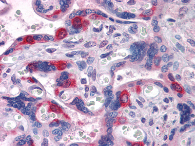
- Immunohistochemistry analysis of paraffin-embedded human Placenta tissues with AEC staining using PEG10 Monoclonal Antibody.
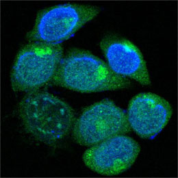
- Confocal immunofluorescence analysis of methanol-fixed HepG2 cells using PEG10 Monoclonal Antibody (green), showing cytoplasmic localization. Blue: DRAQ5 fluorescent DNA dye.

