Oct-3/4 Monoclonal Antibody
- Catalog No.:YM1067
- Applications:WB;FCM;IF
- Reactivity:Human
- Target:
- Oct-3/4
- Fields:
- >>Signaling pathways regulating pluripotency of stem cells
- Gene Name:
- POU5F1
- Protein Name:
- POU domain class 5 transcription factor 1
- Human Gene Id:
- 5460
- Human Swiss Prot No:
- Q01860
- Mouse Swiss Prot No:
- P20263
- Immunogen:
- Purified recombinant human Oct-3/4 protein fragments expressed in E.coli.
- Specificity:
- Oct-3/4 Monoclonal Antibody detects endogenous levels of Oct-3/4 protein.
- Formulation:
- Liquid in PBS containing 50% glycerol, 0.5% BSA and 0.02% sodium azide.
- Source:
- Monoclonal, Mouse
- Dilution:
- WB 1:1000 - 1:2000. Flow cytometry: 1:100 - 1:200. IF 1:100 - 1:500. Not yet tested in other applications.
- Purification:
- Affinity purification
- Concentration:
- 1 mg/ml
- Storage Stability:
- -15°C to -25°C/1 year(Do not lower than -25°C)
- Other Name:
- POU5F1;OCT3;OCT4;OTF3;POU domain; class 5, transcription factor 1;Octamer-binding protein 3;Oct-3;Octamer-binding protein 4;Oct-4;Octamer-binding transcription factor 3;OTF-3
- Molecular Weight(Da):
- 39kD
- Background:
- This gene encodes a transcription factor containing a POU homeodomain that plays a key role in embryonic development and stem cell pluripotency. Aberrant expression of this gene in adult tissues is associated with tumorigenesis. This gene can participate in a translocation with the Ewing's sarcoma gene on chromosome 21, which also leads to tumor formation. Alternative splicing, as well as usage of alternative AUG and non-AUG translation initiation codons, results in multiple isoforms. One of the AUG start codons is polymorphic in human populations. Related pseudogenes have been identified on chromosomes 1, 3, 8, 10, and 12. [provided by RefSeq, Oct 2013],
- Function:
- function:Transcription factor that binds to the octamer motif (5'-ATTTGCAT-3'). Forms a trimeric complex with SOX2 on DNA and controls the expression of a number of genes involved in embryonic development such as YES1, FGF4, UTF1 and ZFP206. Critical for early embryogenesis and for embryonic stem cell pluripotency.,miscellaneous:Several pseudogenes of POU5F1 have been described on chromosomes 1, 3, 8, 10 and 12. 2 of them, localized in chromosomes 8 and 10, are transcribed in cancer tissues but not in normal ones and may be involved in the regulation of POU5F1 gene activity in carcinogenesis.,online information:Oct-4 entry,PTM:Sumoylation enhances the protein stability, DNA binding and transactivation activity. Sumoylation is required for enhanced YES1 expression.,similarity:Belongs to the POU transcription factor family. Class-5 subfamily.,similarity:Contains 1 homeobox DNA-binding doma
- Subcellular Location:
- Cytoplasm. Nucleus. Expressed in a diffuse and slightly punctuate pattern. Colocalizes with MAPK8 and MAPK9 in the nucleus. .
- Expression:
- Expressed in developing brain. Highest levels found in specific cell layers of the cortex, the olfactory bulb, the hippocampus and the cerebellum. Low levels of expression in adult tissues.
Black TiO2-based nanoprobes for T1-weighted MRI-guided photothermal therapy in CD133 high expressed pancreatic cancer stem-like cells. Biomaterials Science Biomater Sci-Uk. 2018 Jul;6(8):2209-2218 WB Human 1 : 2000 PANC-1 cell,SW1990 cell
Wang, Siqi, et al. "Black TiO2 based nanoprobes for T1-weighted MRI guided photothermal therapy in CD133 high expressed pancreatic cancer stem-like cells." Biomaterials Science (2018).
- June 19-2018
- WESTERN IMMUNOBLOTTING PROTOCOL
- June 19-2018
- IMMUNOHISTOCHEMISTRY-PARAFFIN PROTOCOL
- June 19-2018
- IMMUNOFLUORESCENCE PROTOCOL
- September 08-2020
- FLOW-CYTOMEYRT-PROTOCOL
- May 20-2022
- Cell-Based ELISA│解您多样本WB检测之困扰
- July 13-2018
- CELL-BASED-ELISA-PROTOCOL-FOR-ACETYL-PROTEIN
- July 13-2018
- CELL-BASED-ELISA-PROTOCOL-FOR-PHOSPHO-PROTEIN
- July 13-2018
- Antibody-FAQs
- Products Images

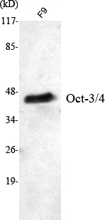
- Western Blot analysis using Oct-3/4 Monoclonal Antibody against F9 cell lysate.

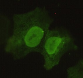
- Immunofluorescence analysis of COS7 cells using Oct-3/4 Monoclonal Antibody.

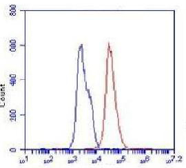
- Flow cytometric analysis of F9 cells stained with Oct-3/4 Monoclonal Antibody (red), followed by FITC-conjugated goat anti-mouse IgG. Blue line histogram represents the isotype control, normal mouse IgG.


