Jak1 mouse mAb
- Catalog No.:YM1316
- Applications:WB;IF;IP
- Reactivity:Human;Rat
- Target:
- JAK1
- Fields:
- >>EGFR tyrosine kinase inhibitor resistance;>>PI3K-Akt signaling pathway;>>Necroptosis;>>Osteoclast differentiation;>>Signaling pathways regulating pluripotency of stem cells;>>NOD-like receptor signaling pathway;>>JAK-STAT signaling pathway;>>Th1 and Th2 cell differentiation;>>Th17 cell differentiation;>>Leishmaniasis;>>Toxoplasmosis;>>Tuberculosis;>>Hepatitis C;>>Hepatitis B;>>Measles;>>Human cytomegalovirus infection;>>Influenza A;>>Human papillomavirus infection;>>Human T-cell leukemia virus 1 infection;>>Kaposi sarcoma-associated herpesvirus infection;>>Herpes simplex virus 1 infection;>>Epstein-Barr virus infection;>>Coronavirus disease - COVID-19;>>Pathways in cancer;>>Viral carcinogenesis;>>Pancreatic cancer;>>PD-L1 expression and PD-1 checkpoint pathway in cancer
- Gene Name:
- jak1
- Human Gene Id:
- 3716
- Human Swiss Prot No:
- P23458
- Mouse Swiss Prot No:
- P52332
- Immunogen:
- Purified recombinant human Jak1 protein fragments expressed in E.coli.
- Specificity:
- This antibody detects endogenous levels of Jak1 and does not cross-react with related proteins.
- Formulation:
- Liquid in PBS containing 50% glycerol, 0.5% BSA and 0.02% sodium azide.
- Source:
- Monoclonal, Mouse
- Dilution:
- wb 1:200-1000 icc 1:200. IF 1:50-200
- Purification:
- The antibody was affinity-purified from mouse ascites by affinity-chromatography using epitope-specific immunogen.
- Concentration:
- 1 mg/ml
- Storage Stability:
- -15°C to -25°C/1 year(Do not lower than -25°C)
- Other Name:
- JAK 1;JAK 1A;JAK 1B;JAK-1;JAK1;JAK1_HUMAN;JAK1A;JAK1B;Janus kinase 1 (a protein tyrosine kinase);Janus kinase 1;JTK3;Tyrosine protein kinase JAK 1;Tyrosine protein kinase JAK1;Tyrosine-protein kinase JAK1.
- Observed Band(KD):
- 130kD
- Background:
- This gene encodes a membrane protein that is a member of a class of protein-tyrosine kinases (PTK) characterized by the presence of a second phosphotransferase-related domain immediately N-terminal to the PTK domain. The encoded kinase phosphorylates STAT proteins (signal transducers and activators of transcription) and plays a key role in interferon-alpha/beta and interferon-gamma signal transduction. Alternative splicing results in multiple transcript variants. [provided by RefSeq, Mar 2016],
- Function:
- catalytic activity:ATP + a [protein]-L-tyrosine = ADP + a [protein]-L-tyrosine phosphate.,domain:Possesses two phosphotransferase domains. The second one probably contains the catalytic domain (By similarity), while the presence of slight differences suggest a different role for domain 1.,domain:The FERM domain mediates interaction with JAKMIP1.,function:Tyrosine kinase of the non-receptor type, involved in the IFN-alpha/beta/gamma signal pathway. Kinase partner for the interleukin (IL)-2 receptor.,sequence caution:Translation N-terminally extended.,similarity:Belongs to the protein kinase superfamily. Tyr protein kinase family. JAK subfamily.,similarity:Contains 1 FERM domain.,similarity:Contains 1 protein kinase domain.,similarity:Contains 1 SH2 domain.,subcellular location:Wholly intracellular, possibly membrane associated.,subunit:Interacts with IL31RA, JAKMIP1 and SHB.,tissue specif
- Subcellular Location:
- Endomembrane system; Peripheral membrane protein. Wholly intracellular, possibly membrane associated.
- Expression:
- Expressed at higher levels in primary colon tumors than in normal colon tissue. The expression level in metastatic colon tumors is comparable to the expression level in normal colon tissue.
- June 19-2018
- WESTERN IMMUNOBLOTTING PROTOCOL
- June 19-2018
- IMMUNOHISTOCHEMISTRY-PARAFFIN PROTOCOL
- June 19-2018
- IMMUNOFLUORESCENCE PROTOCOL
- September 08-2020
- FLOW-CYTOMEYRT-PROTOCOL
- May 20-2022
- Cell-Based ELISA│解您多样本WB检测之困扰
- July 13-2018
- CELL-BASED-ELISA-PROTOCOL-FOR-ACETYL-PROTEIN
- July 13-2018
- CELL-BASED-ELISA-PROTOCOL-FOR-PHOSPHO-PROTEIN
- July 13-2018
- Antibody-FAQs
- Products Images

- Western blot analysis of extracts from C6,Ramos,Jurkat and Hela cell lysates using Jak1 mouse mAb (1:1000 diluted).Predicted band size:130KDa.Observed band size:130KDa.

- Immunocytochemistry staining of HeLa cells fixed with 4% Paraformaldehyde and using anti-Jak1 mouse mAb (dilution 1:200).
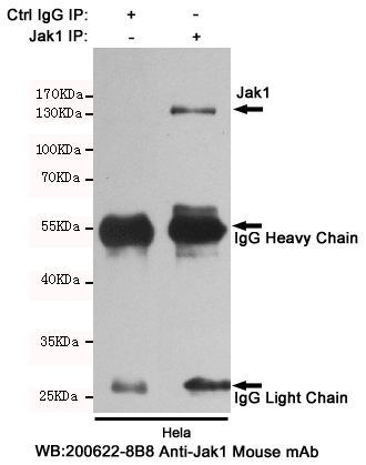
- Immunoprecipitation analysis of Hela cell lysates using Jak1 mouse mAb.


