Kif 7 Monoclonal Antibody(3F8)
- Catalog No.:YM3063
- Applications:IHC;IF
- Reactivity:Human;Mouse;Rat
- Target:
- Kif 7
- Fields:
- >>Hedgehog signaling pathway;>>Pathways in cancer;>>Basal cell carcinoma
- Gene Name:
- KIF7
- Protein Name:
- Kinesin-like protein KIF7
- Human Gene Id:
- 374654
- Human Swiss Prot No:
- Q2M1P5
- Mouse Gene Id:
- 16576
- Mouse Swiss Prot No:
- B7ZNG0
- Immunogen:
- Synthetic Peptide of Kif 7
- Specificity:
- The antibody detects endogenous Kif 7 proteins.
- Formulation:
- PBS, pH 7.4, containing 0.5%BSA, 0.02% sodium azide as Preservative and 50% Glycerol.
- Source:
- Monoclonal, Mouse
- Dilution:
- IHC 1:50-200. IF 1:50-200
- Purification:
- The antibody was affinity-purified from mouse ascites by affinity-chromatography using specific immunogen.
- Storage Stability:
- -15°C to -25°C/1 year(Do not lower than -25°C)
- Other Name:
- Kinesin-like protein KIF7
- Background:
- This gene encodes a cilia-associated protein belonging to the kinesin family. This protein plays a role in the sonic hedgehog (SHH) signaling pathway through the regulation of GLI transcription factors. It functions as a negative regulator of the SHH pathway by preventing inappropriate activation of GLI2 in the absence of ligand, and as a positive regulator by preventing the processing of GLI3 into its repressor form. Mutations in this gene have been associated with various ciliopathies. [provided by RefSeq, Oct 2011],
- Function:
- similarity:Belongs to the kinesin-like protein family. KIF27 subfamily.,similarity:Contains 1 kinesin-motor domain.,tissue specificity:Embryonic stem cells, melanotic melanoma and Jurkat T-cells.,
- Subcellular Location:
- Cell projection, cilium . Cytoplasm, cytoskeleton, cilium basal body . Localizes to the cilium tip.
- Expression:
- Embryonic stem cells, melanotic melanoma and Jurkat T-cells. Expressed in heart, lung, liver, kidney, testis, retina, placenta, pancreas, colon, small intestin, prostate and thymus.
- June 19-2018
- WESTERN IMMUNOBLOTTING PROTOCOL
- June 19-2018
- IMMUNOHISTOCHEMISTRY-PARAFFIN PROTOCOL
- June 19-2018
- IMMUNOFLUORESCENCE PROTOCOL
- September 08-2020
- FLOW-CYTOMEYRT-PROTOCOL
- May 20-2022
- Cell-Based ELISA│解您多样本WB检测之困扰
- July 13-2018
- CELL-BASED-ELISA-PROTOCOL-FOR-ACETYL-PROTEIN
- July 13-2018
- CELL-BASED-ELISA-PROTOCOL-FOR-PHOSPHO-PROTEIN
- July 13-2018
- Antibody-FAQs
- Products Images
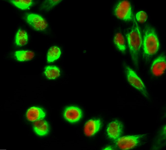
- Immunofluorescence analysis of Hela cell. 1,C/EBP β Polyclonal Antibody(red) was diluted at 1:200(4° overnight). Kif 7 Monoclonal Antibody(3F8)(green) was diluted at 1:200(4° overnight). 2, Goat Anti Rabbit Alexa Fluor 594 Catalog:RS3611 was diluted at 1:1000(room temperature, 50min). Goat Anti Mouse Alexa Fluor 488 Catalog:RS3208 was diluted at 1:1000(room temperature, 50min).

- Immunofluorescence analysis of Mouse-colon tissue. 1,Kif 7 Monoclonal Antibody(3F8)(red) was diluted at 1:200(4°C,overnight). 2, Cy3 labled Secondary antibody was diluted at 1:300(room temperature, 50min).3, Picture B: DAPI(blue) 10min. Picture A:Target. Picture B: DAPI. Picture C: merge of A+B

- IHC staining of Mouse Kidney tissue, diluted at 1:200.
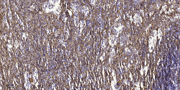
- Immunohistochemical analysis of paraffin-embedded human spleen tissue. 1,primary Antibody was diluted at 1:200(4° overnight). 2, Sodium citrate pH 6.0 was used for antigen retrieval(>98°C,20min). 3,Secondary antibody was diluted at 1:200


