MAP2 Monoclonal Antibody(7D4)
- Catalog No.:YM3067
- Applications:IHC;IF
- Reactivity:Human;Mouse;Rat
- Target:
- MAP-2
- Gene Name:
- MAP2
- Protein Name:
- Microtubule-associated protein 2
- Human Gene Id:
- 4133
- Human Swiss Prot No:
- P11137
- Mouse Swiss Prot No:
- P20357
- Rat Swiss Prot No:
- P15146
- Immunogen:
- Synthetic Peptide of MAP2
- Specificity:
- The antibody detects endogenous MAP2 proteins.
- Formulation:
- PBS, pH 7.4, containing 0.5%BSA, 0.02% sodium azide as Preservative and 50% Glycerol.
- Source:
- Monoclonal, Mouse
- Dilution:
- IHC 1:50-200. IF 1:50-200
- Purification:
- The antibody was affinity-purified from mouse ascites by affinity-chromatography using specific immunogen.
- Storage Stability:
- -15°C to -25°C/1 year(Do not lower than -25°C)
- Other Name:
- MAP2;Microtubule-associated protein 2;MAP-2
- Molecular Weight(Da):
- 200kD
- Background:
- This gene encodes a protein that belongs to the microtubule-associated protein family. The proteins of this family are thought to be involved in microtubule assembly, which is an essential step in neurogenesis. The products of similar genes in rat and mouse are neuron-specific cytoskeletal proteins that are enriched in dentrites, implicating a role in determining and stabilizing dentritic shape during neuron development. A number of alternatively spliced variants encoding distinct isoforms have been described. [provided by RefSeq, Jan 2010],
- Function:
- alternative products:Additional isoforms seem to exist,function:The exact function of MAP2 is unknown but MAPs may stabilize the microtubules against depolymerization. They also seem to have a stiffening effect on microtubules.,PTM:MAP2A/c is phosphorylated. Phosphorylated upon DNA damage, probably by ATM or ATR.,similarity:Contains 3 Tau/MAP repeats.,similarity:Contains 4 Tau/MAP repeats.,
- Subcellular Location:
- Cytoplasm, cytoskeleton . Cell projection, dendrite .
- Expression:
- Brain,Brain cortex,Epithelium,Pancreas,Testis,
- June 19-2018
- WESTERN IMMUNOBLOTTING PROTOCOL
- June 19-2018
- IMMUNOHISTOCHEMISTRY-PARAFFIN PROTOCOL
- June 19-2018
- IMMUNOFLUORESCENCE PROTOCOL
- September 08-2020
- FLOW-CYTOMEYRT-PROTOCOL
- May 20-2022
- Cell-Based ELISA│解您多样本WB检测之困扰
- July 13-2018
- CELL-BASED-ELISA-PROTOCOL-FOR-ACETYL-PROTEIN
- July 13-2018
- CELL-BASED-ELISA-PROTOCOL-FOR-PHOSPHO-PROTEIN
- July 13-2018
- Antibody-FAQs
- Products Images
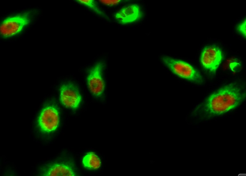
- Immunofluorescence analysis of Hela cell. 1,AQP2 Polyclonal Antibody(green) was diluted at 1:200(4° overnight). (red) was diluted at 1:200(4° overnight). 2, Goat Anti Rabbit Alexa Fluor 488 Catalog:RS3211 was diluted at 1:1000(room temperature, 50min). Goat Anti Mouse Alexa Fluor 594 Catalog:RS3608 was diluted at 1:1000(room temperature, 50min).
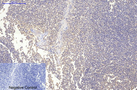
- Immunohistochemical analysis of paraffin-embedded Human-Tonsil tissue. 1,MAP2 Monoclonal Antibody(7D4) was diluted at 1:200(4°C,overnight). 2, Sodium citrate pH 6.0 was used for antibody retrieval(>98°C,20min). 3,Secondary antibody was diluted at 1:200(room tempeRature, 30min). Negative control was used by secondary antibody only.
- Immunohistochemical analysis of paraffin-embedded Rat-kidney tissue. 1,MAP2 Monoclonal Antibody(7D4) was diluted at 1:200(4°C,overnight). 2, Sodium citrate pH 6.0 was used for antibody retrieval(>98°C,20min). 3,Secondary antibody was diluted at 1:200(room tempeRature, 30min). Negative control was used by secondary antibody only.

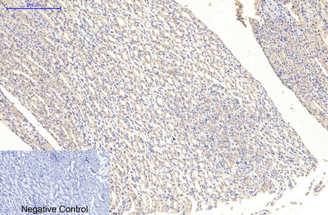
- Immunohistochemical analysis of paraffin-embedded Mouse-brain tissue. 1,MAP2 Monoclonal Antibody(7D4) was diluted at 1:200(4°C,overnight). 2, Sodium citrate pH 6.0 was used for antibody retrieval(>98°C,20min). 3,Secondary antibody was diluted at 1:200(room tempeRature, 30min). Negative control was used by secondary antibody only.
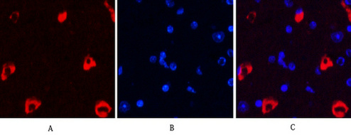
- Immunofluorescence analysis of Mouse-brain tissue. 1,MAP2 Monoclonal Antibody(7D4)(red) was diluted at 1:200(4°C,overnight). 2, Cy3 labled Secondary antibody was diluted at 1:300(room temperature, 50min).3, Picture B: DAPI(blue) 10min. Picture A:Target. Picture B: DAPI. Picture C: merge of A+B
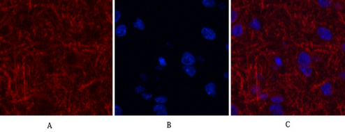
- Immunofluorescence analysis of Rat-brain tissue. 1,MAP2 Monoclonal Antibody(7D4)(red) was diluted at 1:200(4°C,overnight). 2, Cy3 labled Secondary antibody was diluted at 1:300(room temperature, 50min).3, Picture B: DAPI(blue) 10min. Picture A:Target. Picture B: DAPI. Picture C: merge of A+B
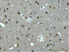
- IHC staining of Human brain tissue paraffin-embedded, diluted at 1:200.
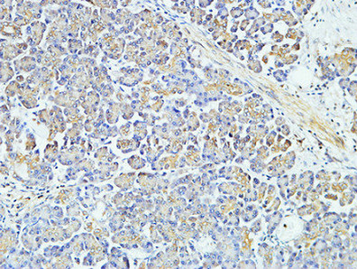
- Immunohistochemical analysis of paraffin-embedded Human pancreas. 1, Antibody was diluted at 1:400(4° overnight). 2, High-pressure and temperature EDTA, pH8.0 was used for antigen retrieval. 3,Secondary antibody was diluted at 1:200(room temperature, 30min).


