Caspase 9 Monoclonal Antibody(3-20)
- Catalog No.:YM3077
- Applications:WB;IHC;IF;IP
- Reactivity:Human;Mouse;Rat;chicken
- Target:
- Caspase-9
- Fields:
- >>Platinum drug resistance;>>p53 signaling pathway;>>PI3K-Akt signaling pathway;>>Apoptosis;>>Apoptosis - multiple species;>>VEGF signaling pathway;>>Thyroid hormone signaling pathway;>>Alzheimer disease;>>Parkinson disease;>>Amyotrophic lateral sclerosis;>>Huntington disease;>>Prion disease;>>Pathways of neurodegeneration - multiple diseases;>>Pathogenic Escherichia coli infection;>>Legionellosis;>>Toxoplasmosis;>>Tuberculosis;>>Hepatitis C;>>Hepatitis B;>>Measles;>>Human cytomegalovirus infection;>>Influenza A;>>Kaposi sarcoma-associated herpesvirus infection;>>Herpes simplex virus 1 infection;>>Epstein-Barr virus infection;>>Human immunodeficiency virus 1 infection;>>Pathways in cancer;>>Colorectal cancer;>>Pancreatic cancer;>>Endometrial cancer;>>Prostate cancer;>>Small cell lung cancer;>>Non-small cell lung cancer;>>Viral myocarditis;>>Lipid and atherosclerosis
- Gene Name:
- CASP9
- Protein Name:
- Caspase9
- Human Gene Id:
- 842
- Human Swiss Prot No:
- P55211
- Immunogen:
- Synthetic Peptide of Caspase 9
- Specificity:
- The antibody detects endogenous Caspase 9 protein.
- Formulation:
- PBS, pH 7.4, containing 0.5%BSA, 0.02% sodium azide as Preservative and 50% Glycerol.
- Source:
- Monoclonal, Mouse
- Dilution:
- WB 1:1000-5000 IP:1:200 IF 1:200 IHC 1:50-300
- Purification:
- The antibody was affinity-purified from mouse ascites by affinity-chromatography using specific immunogen.
- Storage Stability:
- -15°C to -25°C/1 year(Do not lower than -25°C)
- Other Name:
- CASP9;MCH6;Caspase-9;CASP-9;Apoptotic protease Mch-6;Apoptotic protease-activating factor 3;APAF-3;ICE-like apoptotic protease 6;ICE-LAP6
- Observed Band(KD):
- 46kD
- Background:
- CASP9 encodes a member of the cysteine-aspartic acid protease (caspase) family. Sequential activation of caspases plays a central role in the execution-phase of cell apoptosis. Caspases exist as inactive proenzymes which undergo proteolytic processing at conserved aspartic residues to produce two subunits, large and small, that dimerize to form the active enzyme. Caspase 9 can undergo autoproteolytic processing and activation by the apoptosome, a protein complex of cytochrome c and the apoptotic peptidase activating factor 1; this step is thought to be one of the earliest in the caspase activation cascade. Caspase 9 is thought to play a central role in apoptosis and to be a tumor suppressor. Alternative splicing results in multiple transcript variants.
- Function:
- catalytic activity:Strict requirement for an Asp residue at position P1 and with a marked preference for His at position P2. It has a preferred cleavage sequence of Leu-Gly-His-Asp-|-Xaa.,function:Involved in the activation cascade of caspases responsible for apoptosis execution. Binding of caspase-9 to Apaf-1 leads to activation of the protease which then cleaves and activates caspase-3. Proteolytically cleaves poly(ADP-ribose) polymerase (PARP).,function:Isoform 2 lacks activity is an dominant-negative inhibitor of caspase-9.,online information:Caspase-9 entry,PTM:Cleavages at Asp-315 by granzyme B and at Asp-330 by caspase-3 generate the two active subunits. Caspase-8 and -10 can also be involved in these processing events.,similarity:Belongs to the peptidase C14A family.,similarity:Contains 1 CARD domain.,subunit:Heterotetramer that consists of two anti-parallel arranged heterodimers
- Subcellular Location:
- nucleus,mitochondrion,cytosol,apoptosome,
- Expression:
- Ubiquitous, with highest expression in the heart, moderate expression in liver, skeletal muscle, and pancreas. Low levels in all other tissues. Within the heart, specifically expressed in myocytes.
Nuclear factor Nrf2 promotes glycosidase OGG1 expression by activating the AKT pathway to enhance leukemia cell resistance to cytarabine JOURNAL OF BIOLOGICAL CHEMISTRY Qin Shang, Chengyun Pan, Xi Zhang, Tonghua Yang, Tianzhen Hu, Lin Zheng, Shuyun Cao, Cheng Feng, Xiuying Hu, Xiao Chai, Jishi Wang, Qin Fang WB Human U937 cell, THP-1 cell
Comparison of two kinds of Agrocybe cylindracea polysaccharides: structural characteristic and antitumor activity. An-jun Liu WB Human MGC-803 cell
- June 19-2018
- WESTERN IMMUNOBLOTTING PROTOCOL
- June 19-2018
- IMMUNOHISTOCHEMISTRY-PARAFFIN PROTOCOL
- June 19-2018
- IMMUNOFLUORESCENCE PROTOCOL
- September 08-2020
- FLOW-CYTOMEYRT-PROTOCOL
- May 20-2022
- Cell-Based ELISA│解您多样本WB检测之困扰
- July 13-2018
- CELL-BASED-ELISA-PROTOCOL-FOR-ACETYL-PROTEIN
- July 13-2018
- CELL-BASED-ELISA-PROTOCOL-FOR-PHOSPHO-PROTEIN
- July 13-2018
- Antibody-FAQs
- Products Images
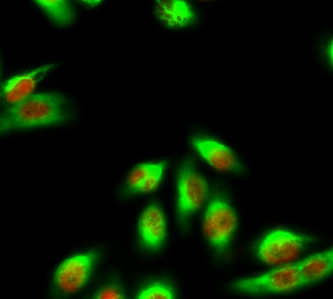
- Immunofluorescence analysis of Hela cell. 1,eIF2α Polyclonal Antibody(red) was diluted at 1:200(4° overnight). Caspase 9 Monoclonal Antibody(3-20)(green) was diluted at 1:200(4° overnight). 2, Goat Anti Rabbit Alexa Fluor 594 Catalog:RS3611 was diluted at 1:1000(room temperature, 50min). Goat Anti Mouse Alexa Fluor 488 Catalog:RS3208 was diluted at 1:1000(room temperature, 50min).

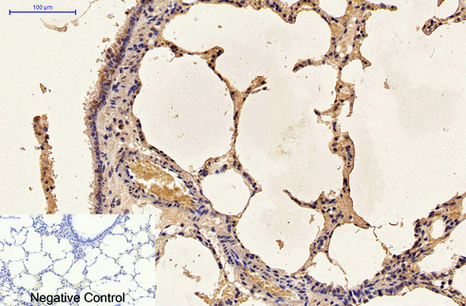
- Immunohistochemical analysis of paraffin-embedded Human-lung tissue. 1,Caspase 9 Monoclonal Antibody(3-20) was diluted at 1:200(4°C,overnight). 2, Sodium citrate pH 6.0 was used for antibody retrieval(>98°C,20min). 3,Secondary antibody was diluted at 1:200(room tempeRature, 30min). Negative control was used by secondary antibody only.
- Immunohistochemical analysis of paraffin-embedded Rat-lung tissue. 1,Caspase 9 Monoclonal Antibody(3-20) was diluted at 1:200(4°C,overnight). 2, Sodium citrate pH 6.0 was used for antibody retrieval(>98°C,20min). 3,Secondary antibody was diluted at 1:200(room tempeRature, 30min). Negative control was used by secondary antibody only.

- Immunohistochemical analysis of paraffin-embedded Mouse-kidney tissue. 1,Caspase 9 Monoclonal Antibody(3-20) was diluted at 1:200(4°C,overnight). 2, Sodium citrate pH 6.0 was used for antibody retrieval(>98°C,20min). 3,Secondary antibody was diluted at 1:200(room tempeRature, 30min). Negative control was used by secondary antibody only.
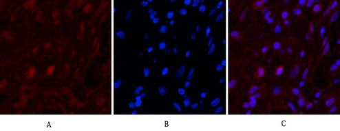
- Immunofluorescence analysis of Human-appendix tissue. 1,Caspase 9 Monoclonal Antibody(3-20)(red) was diluted at 1:200(4°C,overnight). 2, Cy3 labled Secondary antibody was diluted at 1:300(room temperature, 50min).3, Picture B: DAPI(blue) 10min. Picture A:Target. Picture B: DAPI. Picture C: merge of A+B

- Immunofluorescence analysis of Mouse-brain tissue. 1,Caspase 9 Monoclonal Antibody(3-20)(red) was diluted at 1:200(4°C,overnight). 2, Cy3 labled Secondary antibody was diluted at 1:300(room temperature, 50min).3, Picture B: DAPI(blue) 10min. Picture A:Target. Picture B: DAPI. Picture C: merge of A+B

- Immunofluorescence analysis of Rat-spleen tissue. 1,Caspase 9 Monoclonal Antibody(3-20)(red) was diluted at 1:200(4°C,overnight). 2, Cy3 labled Secondary antibody was diluted at 1:300(room temperature, 50min).3, Picture B: DAPI(blue) 10min. Picture A:Target. Picture B: DAPI. Picture C: merge of A+B
- 1) Input: Hela Cell Lysate 2) IP product: IP dilute 1:200
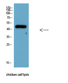
- Western Blot analysis of chicken cell lysis using Antibody diluted at 1:1000
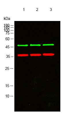
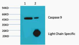
- Western blot analysis of lysates from 1) Hela, 2) Jurkat, 3)3T3 cells, (Green) primary antibody was diluted at 1:1000, 4°over night, secondary antibody(cat:RS23910)was diluted at 1:10000, 37° 1hour. (Red) GAPDH Polyclonal Antibody (cat:YM3215) antibody was diluted at 1:5000 as loading control, 4° over night,secondary antibody(cat:RS23720)was diluted at 1:10000, 37° 1hour.


