EFHD1 Monoclonal Antibody(3G2)
- Catalog No.:YM3085
- Applications:WB;IHC;IF
- Reactivity:Human;Mouse;Rat
- Target:
- EFHD1
- Gene Name:
- EFHD1
- Protein Name:
- EF-hand domain-containing protein D1
- Human Gene Id:
- 80303
- Human Swiss Prot No:
- Q9BUP0
- Mouse Gene Id:
- 98363
- Mouse Swiss Prot No:
- Q9D4J1
- Immunogen:
- Synthetic Peptide of EFHD1
- Specificity:
- The antibody detects endogenous EFHD1 proteins.
- Formulation:
- PBS, pH 7.4, containing 0.5%BSA, 0.02% sodium azide as Preservative and 50% Glycerol.
- Source:
- Monoclonal, Mouse
- Dilution:
- WB 1:2000 IF 1:100-200 IHC 1:50-300
- Purification:
- The antibody was affinity-purified from mouse ascites by affinity-chromatography using specific immunogen.
- Storage Stability:
- -15°C to -25°C/1 year(Do not lower than -25°C)
- Other Name:
- EF-hand domain-containing protein D1 (EF-hand domain-containing protein 1) (Swiprosin-2)
- Observed Band(KD):
- 27kD
- Background:
- This gene encodes a member of the EF-hand super family of calcium binding proteins, which are involved in a variety of cellular processes including mitosis, synaptic transmission, and cytoskeletal rearrangement. The protein encoded by this gene is composed of an N-terminal disordered region, proline-rich elements, two EF-hands, and a C-terminal coiled-coil domain. This protein has been shown to associate with the mitochondrial inner membrane, and in HeLa cells, acts as a novel mitochondrial calcium ion sensor for mitochondrial flash activation. Alternative splicing results in multiple transcript variants. [provided by RefSeq, Jul 2016],
- Function:
- similarity:Contains 2 EF-hand domains.,
- Subcellular Location:
- Mitochondrion inner membrane .
- Expression:
- Brain,Eye,Heart,Hippocampus,Lung,Normal aorta,Placenta,
- June 19-2018
- WESTERN IMMUNOBLOTTING PROTOCOL
- June 19-2018
- IMMUNOHISTOCHEMISTRY-PARAFFIN PROTOCOL
- June 19-2018
- IMMUNOFLUORESCENCE PROTOCOL
- September 08-2020
- FLOW-CYTOMEYRT-PROTOCOL
- May 20-2022
- Cell-Based ELISA│解您多样本WB检测之困扰
- July 13-2018
- CELL-BASED-ELISA-PROTOCOL-FOR-ACETYL-PROTEIN
- July 13-2018
- CELL-BASED-ELISA-PROTOCOL-FOR-PHOSPHO-PROTEIN
- July 13-2018
- Antibody-FAQs
- Products Images
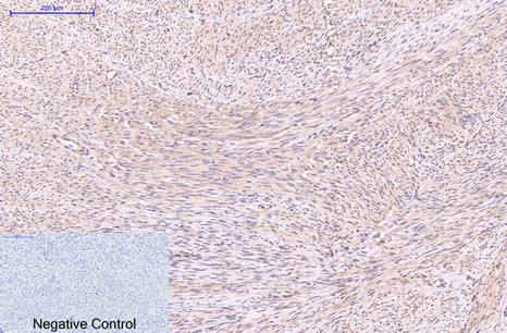
- Immunohistochemical analysis of paraffin-embedded Human-uterus tissue. 1,EFHD1 Monoclonal Antibody(3G2) was diluted at 1:200(4°C,overnight). 2, Sodium citrate pH 6.0 was used for antibody retrieval(>98°C,20min). 3,Secondary antibody was diluted at 1:200(room tempeRature, 30min). Negative control was used by secondary antibody only.
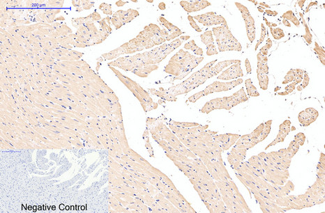
- Immunohistochemical analysis of paraffin-embedded Rat-heart tissue. 1,EFHD1 Monoclonal Antibody(3G2) was diluted at 1:200(4°C,overnight). 2, Sodium citrate pH 6.0 was used for antibody retrieval(>98°C,20min). 3,Secondary antibody was diluted at 1:200(room tempeRature, 30min). Negative control was used by secondary antibody only.

- Immunohistochemical analysis of paraffin-embedded Mouse-testis tissue. 1,EFHD1 Monoclonal Antibody(3G2) was diluted at 1:200(4°C,overnight). 2, Sodium citrate pH 6.0 was used for antibody retrieval(>98°C,20min). 3,Secondary antibody was diluted at 1:200(room tempeRature, 30min). Negative control was used by secondary antibody only.
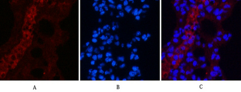
- Immunofluorescence analysis of Mouse-lung tissue. 1,EFHD1 Monoclonal Antibody(3G2)(red) was diluted at 1:200(4°C,overnight). 2, Cy3 labled Secondary antibody was diluted at 1:300(room temperature, 50min).3, Picture B: DAPI(blue) 10min. Picture A:Target. Picture B: DAPI. Picture C: merge of A+B
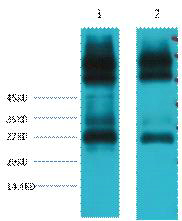
- Western blot analysis of 1) Mouse spleen tissue, 2) Rat spleen tissue, diluted at 1:3000.
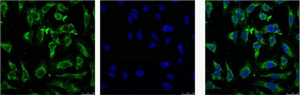
- IF analysis of Hela with antibody (Left) and DAPI (Right) diluted at 1:100.


