ERK1 mouse Monoclonal Antibody(5E9)
- Catalog No.:YM3674
- Applications:IF;WB;IHC
- Reactivity:Human;Rat;Mouse
- Target:
- ERK 1
- Fields:
- >>EGFR tyrosine kinase inhibitor resistance;>>Endocrine resistance;>>Platinum drug resistance;>>MAPK signaling pathway;>>ErbB signaling pathway;>>Ras signaling pathway;>>Rap1 signaling pathway;>>cGMP-PKG signaling pathway;>>cAMP signaling pathway;>>Chemokine signaling pathway;>>HIF-1 signaling pathway;>>FoxO signaling pathway;>>Sphingolipid signaling pathway;>>Phospholipase D signaling pathway;>>Oocyte meiosis;>>Autophagy - animal;>>mTOR signaling pathway;>>PI3K-Akt signaling pathway;>>Apoptosis;>>Cellular senescence;>>Adrenergic signaling in cardiomyocytes;>>Vascular smooth muscle contraction;>>TGF-beta signaling pathway;>>Axon guidance;>>VEGF signaling pathway;>>Apelin signaling pathway;>>Osteoclast differentiation;>>Focal adhesion;>>Adherens junction;>>Gap junction;>>Signaling pathways regulating pluripotency of stem cells;>>Platelet activation;>>Neutrophil extracellular trap formation;>>Toll-like receptor signaling pathway;>>NOD-like receptor signaling pathway;>>C-type lectin recep
- Gene Name:
- MAPK3
- Protein Name:
- MAPK3
- Human Gene Id:
- 5594
- Human Swiss Prot No:
- P27361
- Mouse Swiss Prot No:
- Q63844
- Rat Swiss Prot No:
- P21708
- Immunogen:
- Recombinant Protein of ERK1 of MAPK3
- Specificity:
- ERK1 protein detects endogenous levels of MAPK3
- Formulation:
- Liquid in PBS containing 50% glycerol, 0.5% BSA and 0.02% sodium azide.
- Source:
- Monoclonal, Mouse
- Dilution:
- WB 1:500-2000 IF 1:50-200 IHC 1:100-200
- Purification:
- The antibody was affinity-purified from mouse ascites by affinity-chromatography using specific immunogen.
- Concentration:
- 1 mg/ml
- Storage Stability:
- -15°C to -25°C/1 year(Do not lower than -25°C)
- Other Name:
- MAPK3
- Observed Band(KD):
- 44kD
- Background:
- The protein encoded by this gene is a member of the MAP kinase family. MAP kinases, also known as extracellular signal-regulated kinases (ERKs), act in a signaling cascade that regulates various cellular processes such as proliferation, differentiation, and cell cycle progression in response to a variety of extracellular signals. This kinase is activated by upstream kinases, resulting in its translocation to the nucleus where it phosphorylates nuclear targets. Alternatively spliced transcript variants encoding different protein isoforms have been described. [provided by RefSeq, Jul 2008],
- Function:
- catalytic activity:ATP + a protein = ADP + a phosphoprotein.,cofactor:Magnesium.,domain:The TXY motif contains the threonine and tyrosine residues whose phosphorylation activates the MAP kinases.,enzyme regulation:Activated by tyrosine phosphorylation in response to insulin and NGF.,function:Involved in both the initiation and regulation of meiosis, mitosis, and postmitotic functions in differentiated cells by phosphorylating a number of transcription factors such as ELK-1. Phosphorylates EIF4EBP1; required for initiation of translation. Phosphorylates microtubule-associated protein 2 (MAP2). Phosphorylates SPZ1 (By similarity). Phosphorylates heat shock factor protein 4 (HSF4).,PTM:Dually phosphorylated on Thr-202 and Tyr-204, which activates the enzyme.,similarity:Belongs to the protein kinase superfamily.,similarity:Belongs to the protein kinase superfamily. CMGC Ser/Thr protein kinas
- Subcellular Location:
- Cytoplasm . Nucleus. Membrane, caveola . Cell junction, focal adhesion . Autophosphorylation at Thr-207 promotes nuclear localization (PubMed:19060905). PEA15-binding redirects the biological outcome of MAPK3 kinase-signaling by sequestering MAPK3 into the cytoplasm (By similarity). .
- Expression:
- Epithelium,Eye,Hepatoma,Human cervix,Lymph,
Licoflavone A Suppresses Gastric Cancer Growth and Metastasis by Blocking the VEGFR-2 Signaling Pathway. Journal of Oncology2022;2022:5497991. Human 1 : 1000 MKN-45 cell
- June 19-2018
- WESTERN IMMUNOBLOTTING PROTOCOL
- June 19-2018
- IMMUNOHISTOCHEMISTRY-PARAFFIN PROTOCOL
- June 19-2018
- IMMUNOFLUORESCENCE PROTOCOL
- September 08-2020
- FLOW-CYTOMEYRT-PROTOCOL
- May 20-2022
- Cell-Based ELISA│解您多样本WB检测之困扰
- July 13-2018
- CELL-BASED-ELISA-PROTOCOL-FOR-ACETYL-PROTEIN
- July 13-2018
- CELL-BASED-ELISA-PROTOCOL-FOR-PHOSPHO-PROTEIN
- July 13-2018
- Antibody-FAQs
- Products Images
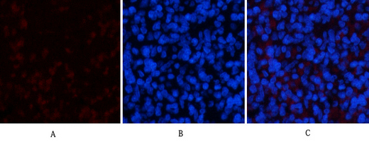
- Immunofluorescence analysis of rat-spleen tissue. 1,ERK1 Mouse Monoclonal Antibody(5E9)(red) was diluted at 1:200(4°C,overnight). 2, Cy3 labled Secondary antibody was diluted at 1:300(room temperature, 50min).3, Picture B: DAPI(blue) 10min. Picture A:Target. Picture B: DAPI. Picture C: merge of A+B
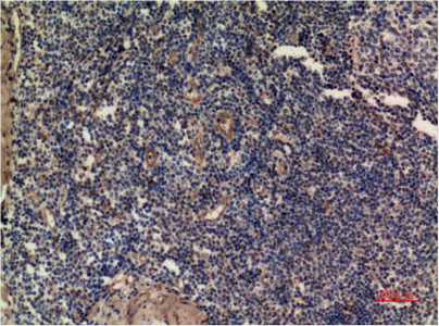
- Immunohistochemical analysis of paraffin-embedded Human Tonsil Tissue using ERK1 Mouse mAb diluted at 1:200.
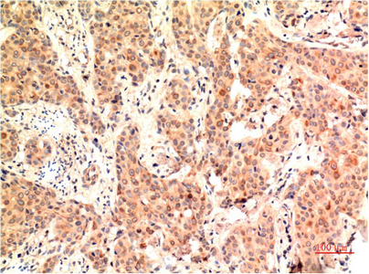
- Immunohistochemical analysis of paraffin-embedded Human Breast Carcinoma Tissue using ERK1 Mouse mAb diluted at 1:200.


