JNK1/2/3 (phospho Thr183) Polyclonal Antibody
- Catalog No.:YP0156
- Applications:WB;IHC;IF;ELISA
- Reactivity:Human;Mouse;Rat;Chicken
- Target:
- JNK1/2/3
- Fields:
- >>Endocrine resistance;>>MAPK signaling pathway;>>ErbB signaling pathway;>>Ras signaling pathway;>>cAMP signaling pathway;>>FoxO signaling pathway;>>Sphingolipid signaling pathway;>>Mitophagy - animal;>>Autophagy - animal;>>Protein processing in endoplasmic reticulum;>>Apoptosis;>>Apoptosis - multiple species;>>Necroptosis;>>Wnt signaling pathway;>>Osteoclast differentiation;>>Focal adhesion;>>Tight junction;>>Toll-like receptor signaling pathway;>>NOD-like receptor signaling pathway;>>RIG-I-like receptor signaling pathway;>>C-type lectin receptor signaling pathway;>>IL-17 signaling pathway;>>Th1 and Th2 cell differentiation;>>Th17 cell differentiation;>>T cell receptor signaling pathway;>>Fc epsilon RI signaling pathway;>>TNF signaling pathway;>>Neurotrophin signaling pathway;>>Retrograde endocannabinoid signaling;>>Dopaminergic synapse;>>Inflammatory mediator regulation of TRP channels;>>Insulin signaling pathway;>>GnRH signaling pathway;>>Progesterone-mediated oocyte maturation;>>Pr
- Gene Name:
- MAPK8/9/10
- Protein Name:
- Mitogen-activated protein kinase 8/9/10
- Human Gene Id:
- 5599/5601/5602
- Human Swiss Prot No:
- P45983/P45984/P53779
- Mouse Gene Id:
- 26419/26420
- Rat Gene Id:
- 116554/50658/25272
- Rat Swiss Prot No:
- P49185/P49186/P49187
- Immunogen:
- The antiserum was produced against synthesized peptide derived from human SAPK/JNK around the phosphorylation site of Thr183. AA range:151-200
- Specificity:
- Phospho-JNK1/2/3 (T183) Polyclonal Antibody detects endogenous levels of JNK1/2/3 protein only when phosphorylated at T183.
- Formulation:
- Liquid in PBS containing 50% glycerol, 0.5% BSA and 0.02% sodium azide.
- Source:
- Polyclonal, Rabbit,IgG
- Dilution:
- WB 1:500 - 1:2000. IHC 1:100 - 1:300. IF 1:200 - 1:1000. ELISA: 1:5000. Not yet tested in other applications.
- Purification:
- The antibody was affinity-purified from rabbit antiserum by affinity-chromatography using epitope-specific immunogen.
- Concentration:
- 1 mg/ml
- Storage Stability:
- -15°C to -25°C/1 year(Do not lower than -25°C)
- Other Name:
- MAPK8;JNK1;PRKM8;SAPK1;SAPK1C;Mitogen-activated protein kinase 8;MAP kinase 8;MAPK 8;JNK-46;Stress-activated protein kinase 1c;SAPK1c;Stress-activated protein kinase JNK1;c-Jun N-terminal kinase 1;MAPK9;JNK2;PRKM9;SAPK1A;Mi
- Observed Band(KD):
- 46kD,54kD
- Background:
- The protein encoded by this gene is a member of the MAP kinase family. MAP kinases act as an integration point for multiple biochemical signals, and are involved in a wide variety of cellular processes such as proliferation, differentiation, transcription regulation and development. This kinase is activated by various cell stimuli, and targets specific transcription factors, and thus mediates immediate-early gene expression in response to cell stimuli. The activation of this kinase by tumor-necrosis factor alpha (TNF-alpha) is found to be required for TNF-alpha induced apoptosis. This kinase is also involved in UV radiation induced apoptosis, which is thought to be related to cytochrom c-mediated cell death pathway. Studies of the mouse counterpart of this gene suggested that this kinase play a key role in T cell proliferation, apoptosis and differentiation. Several alternatively spl
- Function:
- catalytic activity:ATP + a protein = ADP + a phosphoprotein.,cofactor:Magnesium.,domain:The TXY motif contains the threonine and tyrosine residues whose phosphorylation activates the MAP kinases.,enzyme regulation:Activated by threonine and tyrosine phosphorylation by either of two dual specificity kinases, MAP2K4 and MAP2K7. Inhibited by dual specificity phosphatases, such as DUSP1.,function:JNK1 isoforms display different binding patterns: beta-1 preferentially binds to c-Jun, whereas alpha-1, alpha-2, and beta-2 have a similar low level of binding to both c-Jun or ATF2. However, there is no correlation between binding and phosphorylation, which is achieved at about the same efficiency by all isoforms.,function:Responds to activation by environmental stress and pro-inflammatory cytokines by phosphorylating a number of transcription factors, primarily components of AP-1 such as JUN, JDP
- Subcellular Location:
- Cytoplasm . Nucleus . Cell junction, synapse . In the cortical neurons, predominantly cytoplasmic and associated with the Golgi apparatus and endosomal fraction. Increased neuronal activity increases phosphorylated form at synapses (By similarity). Colocalizes with POU5F1 in the nucleus. .
- Expression:
- Brain,Epithelium,Fetal brain,Lung,Pooled,Testis,
GCN5 participates in KLF4-VEGFA feedback to promote endometrial angiogenesis WB Human /HEMECs
Tamoxifen induces the development of hernia in mice by activating MMP-2 and MMP-13 expression. BIOCHIMICA ET BIOPHYSICA ACTA-MOLECULAR BASIS OF DISEASE 2015 Feb 19 WB Mouse NIH3T3 cell
The Effects of Maternal Atrazine Exposure and Swimming Training on Spatial Learning Memory and Hippocampal Morphology in Offspring Male Rats via PSD95/NR2B Signaling Pathway. CELLULAR AND MOLECULAR NEUROBIOLOGY Cell Mol Neurobiol. 2019 Oct;39(7):1003-1015 WB Rat 1:1000 hippocampus
The Role and Mechanism of SIRT1 in Resveratrol-regulated Osteoblast Autophagy in Osteoporosis Rats. Scientific Reports Sci Rep-Uk. 2019 Dec;9(1):1-15 WB Mouse osteoblasts
Inhibiting the Piezo1 channel protects microglia from acute hyperglycaemia damage through the JNK1 and mTOR signalling pathways. LIFE SCIENCES Life Sci. 2021 Jan;264:118667 WB Mouse BV2 cell
Telekin suppresses human hepatocellular carcinoma cells in vitro by inducing G 2 /M phase arrest via the p38 MAPK signaling pathway. ACTA PHARMACOLOGICA SINICA Acta Pharmacol Sin. 2014 Sep;35(10):1311-1322 WB Human HepG2 cell
Luo, Heguo, et al. "Yoda1 Activates Piezo1 in Vitro to Simulate the Upregulation of Piezo1 in the Infected Brain: Piezo1 Participates in the Immune Activation of Microglia." (2021).
Cadmium induces BNIP3-dependent autophagy in chicken spleen by modulating miR-33-AMPK axis. CHEMOSPHERE 2017 Dec 06 WB Chicken 1:1000 Spleen
Alpha-Momorcharin Inhibits Proinflammatory Cytokine Expression by M1 Macrophages but Not Anti-Inflammatory Cytokine Expression by M2 Macrophages Journal of Inflammation Research Fubing Shen WB Human
Morroniside Protects Human Granulosa Cells against H2O2-Induced Oxidative Damage by Regulating the Nrf2 and MAPK Signaling Pathways Evidence-based Complementary and Alternative Medicine Huilan Du WB Human
- June 19-2018
- WESTERN IMMUNOBLOTTING PROTOCOL
- June 19-2018
- IMMUNOHISTOCHEMISTRY-PARAFFIN PROTOCOL
- June 19-2018
- IMMUNOFLUORESCENCE PROTOCOL
- September 08-2020
- FLOW-CYTOMEYRT-PROTOCOL
- May 20-2022
- Cell-Based ELISA│解您多样本WB检测之困扰
- July 13-2018
- CELL-BASED-ELISA-PROTOCOL-FOR-ACETYL-PROTEIN
- July 13-2018
- CELL-BASED-ELISA-PROTOCOL-FOR-PHOSPHO-PROTEIN
- July 13-2018
- Antibody-FAQs
- Products Images
.jpg)
- Jiao, Zhihui, et al. "Adipose-derived stem cells protect ischemia-reperfusion and partial hepatectomy by attenuating endoplasmic reticulum stress." Frontiers in cell and developmental biology 8 (2020): 177.
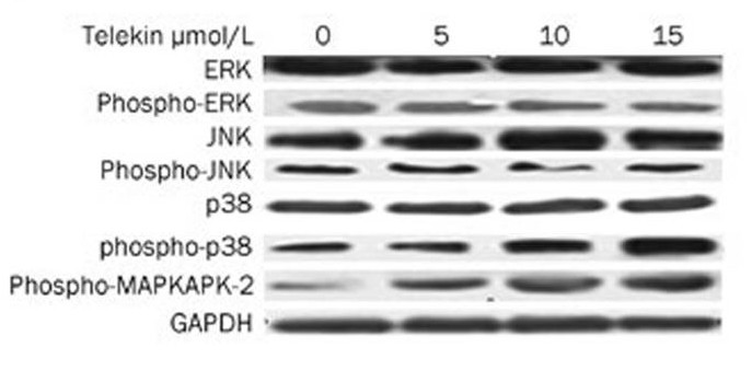
- Li, Lin, et al. "Telekin suppresses human hepatocellular carcinoma cells in vitro by inducing G 2/M phase arrest via the p38 MAPK signaling pathway." Acta Pharmacologica Sinica 35.10 (2014): 1311.
-if-62.jpg)
- Immunofluorescence analysis of rat-testis tissue. 1,JNK1/2/3 (phospho Thr183) Polyclonal Antibody(red) was diluted at 1:200(4°C,overnight). 2, Cy3 labled Secondary antibody was diluted at 1:300(room temperature, 50min).3, Picture B: DAPI(blue) 10min. Picture A:Target. Picture B: DAPI. Picture C: merge of A+B
-if-63.jpg)
- Immunofluorescence analysis of rat-testis tissue. 1,JNK1/2/3 (phospho Thr183) Polyclonal Antibody(red) was diluted at 1:200(4°C,overnight). 2, Cy3 labled Secondary antibody was diluted at 1:300(room temperature, 50min).3, Picture B: DAPI(blue) 10min. Picture A:Target. Picture B: DAPI. Picture C: merge of A+B
-if-64.jpg)
- Immunofluorescence analysis of rat-liver tissue. 1,JNK1/2/3 (phospho Thr183) Polyclonal Antibody(red) was diluted at 1:200(4°C,overnight). 2, Cy3 labled Secondary antibody was diluted at 1:300(room temperature, 50min).3, Picture B: DAPI(blue) 10min. Picture A:Target. Picture B: DAPI. Picture C: merge of A+B
-if-65.jpg)
- Immunofluorescence analysis of rat-liver tissue. 1,JNK1/2/3 (phospho Thr183) Polyclonal Antibody(red) was diluted at 1:200(4°C,overnight). 2, Cy3 labled Secondary antibody was diluted at 1:300(room temperature, 50min).3, Picture B: DAPI(blue) 10min. Picture A:Target. Picture B: DAPI. Picture C: merge of A+B
poly-ihc-rat-kidney.jpg)
- Immunohistochemical analysis of paraffin-embedded Rat-kidney tissue. 1,JNK1/2/3 (phospho Thr183) Polyclonal Antibody was diluted at 1:200(4°C,overnight). 2, Sodium citrate pH 6.0 was used for antibody retrieval(>98°C,20min). 3,Secondary antibody was diluted at 1:200(room tempeRature, 30min). Negative control was used by secondary antibody only.
poly-ihc-human-uterus.jpg)
- Immunohistochemical analysis of paraffin-embedded Human-uterus tissue. 1,JNK1/2/3 (phospho Thr183) Polyclonal Antibody was diluted at 1:200(4°C,overnight). 2, Sodium citrate pH 6.0 was used for antibody retrieval(>98°C,20min). 3,Secondary antibody was diluted at 1:200(room tempeRature, 30min). Negative control was used by secondary antibody only.
poly-ihc-human-uterus-cancer.jpg)
- Immunohistochemical analysis of paraffin-embedded Human-uterus-cancer tissue. 1,JNK1/2/3 (phospho Thr183) Polyclonal Antibody was diluted at 1:200(4°C,overnight). 2, Sodium citrate pH 6.0 was used for antibody retrieval(>98°C,20min). 3,Secondary antibody was diluted at 1:200(room tempeRature, 30min). Negative control was used by secondary antibody only.
poly-ihc-human-colon.jpg)
- Immunohistochemical analysis of paraffin-embedded Human-colon tissue. 1,JNK1/2/3 (phospho Thr183) Polyclonal Antibody was diluted at 1:200(4°C,overnight). 2, Sodium citrate pH 6.0 was used for antibody retrieval(>98°C,20min). 3,Secondary antibody was diluted at 1:200(room tempeRature, 30min). Negative control was used by secondary antibody only.
poly-ihc-human-liver.jpg)
- Immunohistochemical analysis of paraffin-embedded Human-liver tissue. 1,JNK1/2/3 (phospho Thr183) Polyclonal Antibody was diluted at 1:200(4°C,overnight). 2, Sodium citrate pH 6.0 was used for antibody retrieval(>98°C,20min). 3,Secondary antibody was diluted at 1:200(room tempeRature, 30min). Negative control was used by secondary antibody only.
poly-ihc-rat-spleen.jpg)
- Immunohistochemical analysis of paraffin-embedded Rat-spleen tissue. 1,JNK1/2/3 (phospho Thr183) Polyclonal Antibody was diluted at 1:200(4°C,overnight). 2, Sodium citrate pH 6.0 was used for antibody retrieval(>98°C,20min). 3,Secondary antibody was diluted at 1:200(room tempeRature, 30min). Negative control was used by secondary antibody only.
poly-ihc-mouse-heart.jpg)
- Immunohistochemical analysis of paraffin-embedded Mouse-heart tissue. 1,JNK1/2/3 (phospho Thr183) Polyclonal Antibody was diluted at 1:200(4°C,overnight). 2, Sodium citrate pH 6.0 was used for antibody retrieval(>98°C,20min). 3,Secondary antibody was diluted at 1:200(room tempeRature, 30min). Negative control was used by secondary antibody only.
poly-ihc-mouse-testis.jpg)
- Immunohistochemical analysis of paraffin-embedded Mouse-testis tissue. 1,JNK1/2/3 (phospho Thr183) Polyclonal Antibody was diluted at 1:200(4°C,overnight). 2, Sodium citrate pH 6.0 was used for antibody retrieval(>98°C,20min). 3,Secondary antibody was diluted at 1:200(room tempeRature, 30min). Negative control was used by secondary antibody only.
poly-ihc-mouse-colon.jpg)
- Immunohistochemical analysis of paraffin-embedded Mouse-colon tissue. 1,JNK1/2/3 (phospho Thr183) Polyclonal Antibody was diluted at 1:200(4°C,overnight). 2, Sodium citrate pH 6.0 was used for antibody retrieval(>98°C,20min). 3,Secondary antibody was diluted at 1:200(room tempeRature, 30min). Negative control was used by secondary antibody only.
poly-ihc-mouse-kidney.jpg)
- Immunohistochemical analysis of paraffin-embedded Mouse-kidney tissue. 1,JNK1/2/3 (phospho Thr183) Polyclonal Antibody was diluted at 1:200(4°C,overnight). 2, Sodium citrate pH 6.0 was used for antibody retrieval(>98°C,20min). 3,Secondary antibody was diluted at 1:200(room tempeRature, 30min). Negative control was used by secondary antibody only.
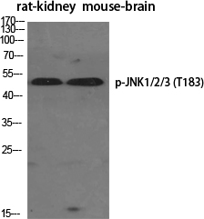
- Western Blot analysis of various cells using Phospho-JNK1/2/3 (T183) Polyclonal Antibody diluted at 1:1000
.jpg)
- Western Blot analysis of MCF-7 cells using Phospho-JNK1/2/3 (T183) Polyclonal Antibody diluted at 1:1000
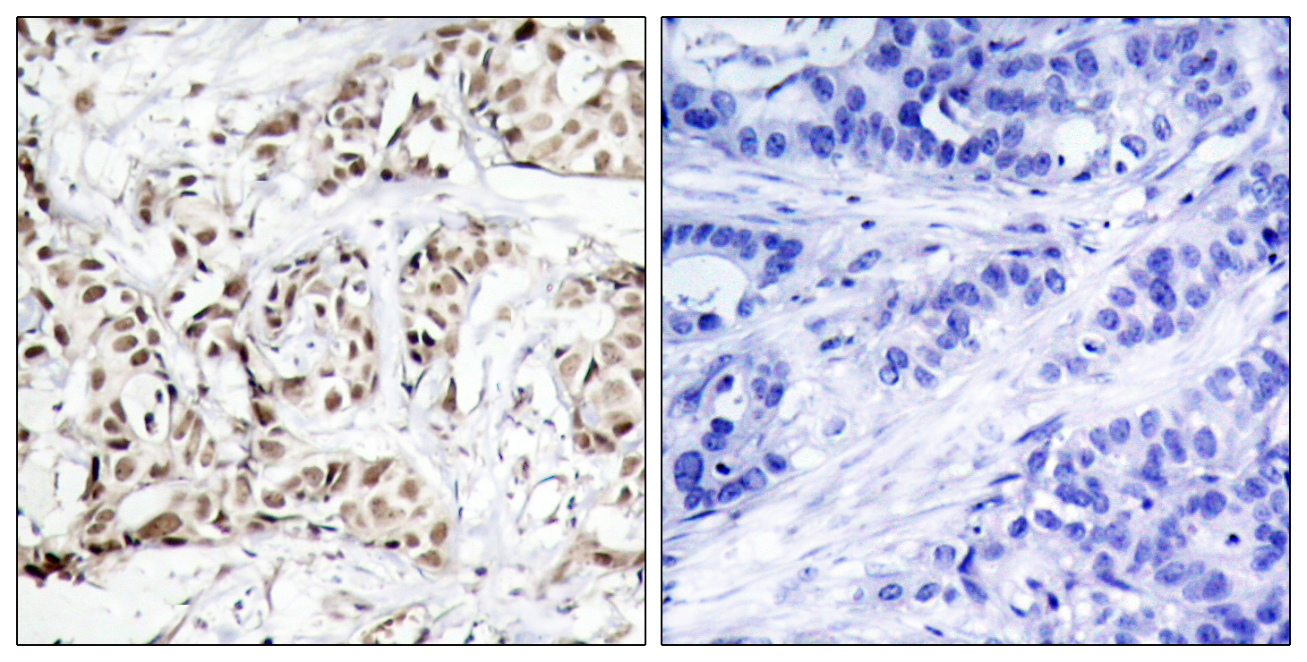
- Immunohistochemistry analysis of paraffin-embedded human breast carcinoma, using SAPK/JNK (Phospho-Thr183) Antibody. The picture on the right is blocked with the phospho peptide.
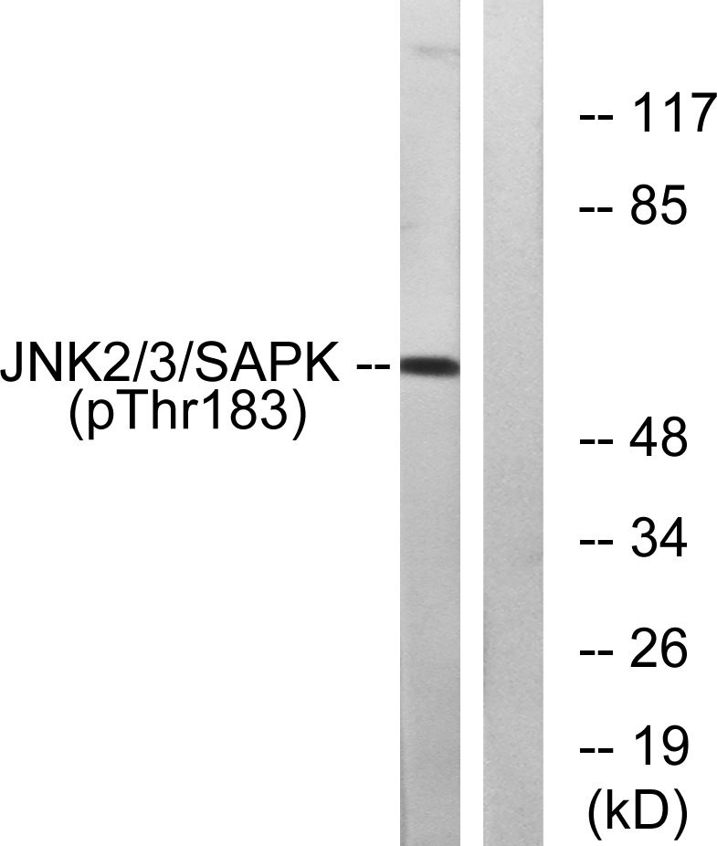
- Western blot analysis of lysates from HeLa cells treated with Anisomycin 200ng/ml 10', using SAPK/JNK (Phospho-Thr183) Antibody. The lane on the right is blocked with the phospho peptide.


