NFκB-p65 (phospho Ser529) Polyclonal Antibody
- Catalog No.:YP0189
- Applications:IF;WB;IHC;ELISA
- Reactivity:Human;Mouse;Rat;Monkey
- Target:
- NFkB p65
- Fields:
- >>Antifolate resistance;>>MAPK signaling pathway;>>Ras signaling pathway;>>cAMP signaling pathway;>>Chemokine signaling pathway;>>NF-kappa B signaling pathway;>>HIF-1 signaling pathway;>>Sphingolipid signaling pathway;>>Mitophagy - animal;>>PI3K-Akt signaling pathway;>>Apoptosis;>>Longevity regulating pathway;>>Cellular senescence;>>Osteoclast differentiation;>>Neutrophil extracellular trap formation;>>Toll-like receptor signaling pathway;>>NOD-like receptor signaling pathway;>>RIG-I-like receptor signaling pathway;>>Cytosolic DNA-sensing pathway;>>C-type lectin receptor signaling pathway;>>IL-17 signaling pathway;>>Th1 and Th2 cell differentiation;>>Th17 cell differentiation;>>T cell receptor signaling pathway;>>B cell receptor signaling pathway;>>TNF signaling pathway;>>Neurotrophin signaling pathway;>>Prolactin signaling pathway;>>Adipocytokine signaling pathway;>>Relaxin signaling pathway;>>Insulin resistance;>>Non-alcoholic fatty liver disease;>>AGE-RAGE signaling pathway in diabe
- Gene Name:
- RELA
- Protein Name:
- Transcription factor p65
- Human Gene Id:
- 5970
- Human Swiss Prot No:
- Q04206
- Mouse Gene Id:
- 19697
- Mouse Swiss Prot No:
- Q04207
- Immunogen:
- Synthesized phospho-peptide around the phosphorylation site of human NFκB-p65 (phospho Ser529)
- Specificity:
- Phospho-NFκB-p65 (S529) Polyclonal Antibody detects endogenous levels of NFκB-p65 protein only when phosphorylated at S529.
- Formulation:
- Liquid in PBS containing 50% glycerol, 0.5% BSA and 0.02% sodium azide.
- Source:
- Polyclonal, Rabbit,IgG
- Dilution:
- IF 1:50-200 WB 1:500 - 1:2000. IHC 1:100 - 1:300. ELISA: 1:20000. Not yet tested in other applications.
- Purification:
- The antibody was affinity-purified from rabbit antiserum by affinity-chromatography using epitope-specific immunogen.
- Concentration:
- 1 mg/ml
- Storage Stability:
- -15°C to -25°C/1 year(Do not lower than -25°C)
- Other Name:
- RELA;NFKB3;Transcription factor p65;Nuclear factor NF-kappa-B p65 subunit;Nuclear factor of kappa light polypeptide gene enhancer in B-cells 3
- Observed Band(KD):
- 60kD
- Background:
- NF-kappa-B is a ubiquitous transcription factor involved in several biological processes. It is held in the cytoplasm in an inactive state by specific inhibitors. Upon degradation of the inhibitor, NF-kappa-B moves to the nucleus and activates transcription of specific genes. NF-kappa-B is composed of NFKB1 or NFKB2 bound to either REL, RELA, or RELB. The most abundant form of NF-kappa-B is NFKB1 complexed with the product of this gene, RELA. Four transcript variants encoding different isoforms have been found for this gene. [provided by RefSeq, Sep 2011],
- Function:
- function:NF-kappa-B is a pleiotropic transcription factor which is present in almost all cell types and is involved in many biological processed such as inflammation, immunity, differentiation, cell growth, tumorigenesis and apoptosis. NF-kappa-B is a homo- or heterodimeric complex formed by the Rel-like domain-containing proteins RELA/p65, RELB, NFKB1/p105, NFKB1/p50, REL and NFKB2/p52 and the heterodimeric p65-p50 complex appears to be most abundant one. The dimers bind at kappa-B sites in the DNA of their target genes and the individual dimers have distinct preferences for different kappa-B sites that they can bind with distinguishable affinity and specificity. Different dimer combinations act as transcriptional activators or repressors, respectively. NF-kappa-B is controlled by various mechanisms of post-translational modification and subcellular compartmentalization as well as by in
- Subcellular Location:
- Nucleus . Cytoplasm . Nuclear, but also found in the cytoplasm in an inactive form complexed to an inhibitor (I-kappa-B) (PubMed:1493333). Colocalized with DDX1 in the nucleus upon TNF-alpha induction (PubMed:19058135). Colocalizes with GFI1 in the nucleus after LPS stimulation (PubMed:20547752). Translocation to the nucleus is impaired in L.monocytogenes infection (PubMed:20855622). .
- Expression:
- Bone,Colon,Pancreas,Placenta,
Effect of the Pulsatilla decoction n-butanol extract on vulvovaginal candidiasis caused by Candida glabrata and on its virulence factors FITOTERAPIA Jiaping Zhang WB Mouse vaginal tissue
Polystyrene nanoplastics induce intestinal and hepatic inflammation through activation of NF-κB/NLRP3 pathways and related gut-liver axis in mice SCIENCE OF THE TOTAL ENVIRONMENT Xuanwei Chen WB Mouse 1:1000 intestinal tissue,liver tissue
POSTN knockdown suppresses IL-1β-induced inflammation and apoptosis of nucleus pulposus cells via inhibiting the NF-κB pathway and alleviates intervertebral disc degeneration Journal of Cell Communication and Signaling Zhaoheng Wang WB Rat 1:1000 nucleus pulposus cell (NPC)
- June 19-2018
- WESTERN IMMUNOBLOTTING PROTOCOL
- June 19-2018
- IMMUNOHISTOCHEMISTRY-PARAFFIN PROTOCOL
- June 19-2018
- IMMUNOFLUORESCENCE PROTOCOL
- September 08-2020
- FLOW-CYTOMEYRT-PROTOCOL
- May 20-2022
- Cell-Based ELISA│解您多样本WB检测之困扰
- July 13-2018
- CELL-BASED-ELISA-PROTOCOL-FOR-ACETYL-PROTEIN
- July 13-2018
- CELL-BASED-ELISA-PROTOCOL-FOR-PHOSPHO-PROTEIN
- July 13-2018
- Antibody-FAQs
- Products Images
-if-74.jpg)
- Immunofluorescence analysis of human-lung tissue. 1,NFκB-p65 (phospho Ser529) Polyclonal Antibody(red) was diluted at 1:200(4°C,overnight). 2, Cy3 labled Secondary antibody was diluted at 1:300(room temperature, 50min).3, Picture B: DAPI(blue) 10min. Picture A:Target. Picture B: DAPI. Picture C: merge of A+B
-if-75.jpg)
- Immunofluorescence analysis of human-lung tissue. 1,NFκB-p65 (phospho Ser529) Polyclonal Antibody(red) was diluted at 1:200(4°C,overnight). 2, Cy3 labled Secondary antibody was diluted at 1:300(room temperature, 50min).3, Picture B: DAPI(blue) 10min. Picture A:Target. Picture B: DAPI. Picture C: merge of A+B
-if-76.jpg)
- Immunofluorescence analysis of rat-heart tissue. 1,NFκB-p65 (phospho Ser529) Polyclonal Antibody(red) was diluted at 1:200(4°C,overnight). 2, Cy3 labled Secondary antibody was diluted at 1:300(room temperature, 50min).3, Picture B: DAPI(blue) 10min. Picture A:Target. Picture B: DAPI. Picture C: merge of A+B
-if-77.jpg)
- Immunofluorescence analysis of rat-heart tissue. 1,NFκB-p65 (phospho Ser529) Polyclonal Antibody(red) was diluted at 1:200(4°C,overnight). 2, Cy3 labled Secondary antibody was diluted at 1:300(room temperature, 50min).3, Picture B: DAPI(blue) 10min. Picture A:Target. Picture B: DAPI. Picture C: merge of A+B
-if-78.jpg)
- Immunofluorescence analysis of mouse-spleen tissue. 1,NFκB-p65 (phospho Ser529) Polyclonal Antibody(red) was diluted at 1:200(4°C,overnight). 2, Cy3 labled Secondary antibody was diluted at 1:300(room temperature, 50min).3, Picture B: DAPI(blue) 10min. Picture A:Target. Picture B: DAPI. Picture C: merge of A+B
-if-79.jpg)
- Immunofluorescence analysis of mouse-spleen tissue. 1,NFκB-p65 (phospho Ser529) Polyclonal Antibody(red) was diluted at 1:200(4°C,overnight). 2, Cy3 labled Secondary antibody was diluted at 1:300(room temperature, 50min).3, Picture B: DAPI(blue) 10min. Picture A:Target. Picture B: DAPI. Picture C: merge of A+B
poly-ihc-human-uterus.jpg)
- Immunohistochemical analysis of paraffin-embedded Human-uterus tissue. 1,NFκB-p65 (phospho Ser529) Polyclonal Antibody was diluted at 1:200(4°C,overnight). 2, Sodium citrate pH 6.0 was used for antibody retrieval(>98°C,20min). 3,Secondary antibody was diluted at 1:200(room tempeRature, 30min). Negative control was used by secondary antibody only.
poly-ihc-human-uterus-cancer.jpg)
- Immunohistochemical analysis of paraffin-embedded Human-uterus-cancer tissue. 1,NFκB-p65 (phospho Ser529) Polyclonal Antibody was diluted at 1:200(4°C,overnight). 2, Sodium citrate pH 6.0 was used for antibody retrieval(>98°C,20min). 3,Secondary antibody was diluted at 1:200(room tempeRature, 30min). Negative control was used by secondary antibody only.
poly-ihc-human-lung-cancer.jpg)
- Immunohistochemical analysis of paraffin-embedded Human-lung-cancer tissue. 1,NFκB-p65 (phospho Ser529) Polyclonal Antibody was diluted at 1:200(4°C,overnight). 2, Sodium citrate pH 6.0 was used for antibody retrieval(>98°C,20min). 3,Secondary antibody was diluted at 1:200(room tempeRature, 30min). Negative control was used by secondary antibody only.
poly-ihc-rat-heart.jpg)
- Immunohistochemical analysis of paraffin-embedded Rat-heart tissue. 1,NFκB-p65 (phospho Ser529) Polyclonal Antibody was diluted at 1:200(4°C,overnight). 2, Sodium citrate pH 6.0 was used for antibody retrieval(>98°C,20min). 3,Secondary antibody was diluted at 1:200(room tempeRature, 30min). Negative control was used by secondary antibody only.
poly-ihc-rat-liver.jpg)
- Immunohistochemical analysis of paraffin-embedded Rat-liver tissue. 1,NFκB-p65 (phospho Ser529) Polyclonal Antibody was diluted at 1:200(4°C,overnight). 2, Sodium citrate pH 6.0 was used for antibody retrieval(>98°C,20min). 3,Secondary antibody was diluted at 1:200(room tempeRature, 30min). Negative control was used by secondary antibody only.
poly-ihc-rat-kidney.jpg)
- Immunohistochemical analysis of paraffin-embedded Rat-kidney tissue. 1,NFκB-p65 (phospho Ser529) Polyclonal Antibody was diluted at 1:200(4°C,overnight). 2, Sodium citrate pH 6.0 was used for antibody retrieval(>98°C,20min). 3,Secondary antibody was diluted at 1:200(room tempeRature, 30min). Negative control was used by secondary antibody only.
poly-ihc-rat-spinal-cord.jpg)
- Immunohistochemical analysis of paraffin-embedded Rat-spinal-cord tissue. 1,NFκB-p65 (phospho Ser529) Polyclonal Antibody was diluted at 1:200(4°C,overnight). 2, Sodium citrate pH 6.0 was used for antibody retrieval(>98°C,20min). 3,Secondary antibody was diluted at 1:200(room tempeRature, 30min). Negative control was used by secondary antibody only.
poly-ihc-rat-brain.jpg)
- Immunohistochemical analysis of paraffin-embedded Rat-brain tissue. 1,NFκB-p65 (phospho Ser529) Polyclonal Antibody was diluted at 1:200(4°C,overnight). 2, Sodium citrate pH 6.0 was used for antibody retrieval(>98°C,20min). 3,Secondary antibody was diluted at 1:200(room tempeRature, 30min). Negative control was used by secondary antibody only.
poly-ihc-rat-spleen.jpg)
- Immunohistochemical analysis of paraffin-embedded Rat-spleen tissue. 1,NFκB-p65 (phospho Ser529) Polyclonal Antibody was diluted at 1:200(4°C,overnight). 2, Sodium citrate pH 6.0 was used for antibody retrieval(>98°C,20min). 3,Secondary antibody was diluted at 1:200(room tempeRature, 30min). Negative control was used by secondary antibody only.
poly-ihc-mouse-liver.jpg)
- Immunohistochemical analysis of paraffin-embedded Mouse-liver tissue. 1,NFκB-p65 (phospho Ser529) Polyclonal Antibody was diluted at 1:200(4°C,overnight). 2, Sodium citrate pH 6.0 was used for antibody retrieval(>98°C,20min). 3,Secondary antibody was diluted at 1:200(room tempeRature, 30min). Negative control was used by secondary antibody only.
poly-ihc-mouse-lung.jpg)
- Immunohistochemical analysis of paraffin-embedded Mouse-lung tissue. 1,NFκB-p65 (phospho Ser529) Polyclonal Antibody was diluted at 1:200(4°C,overnight). 2, Sodium citrate pH 6.0 was used for antibody retrieval(>98°C,20min). 3,Secondary antibody was diluted at 1:200(room tempeRature, 30min). Negative control was used by secondary antibody only.
poly-ihc-mouse-kidney.jpg)
- Immunohistochemical analysis of paraffin-embedded Mouse-kidney tissue. 1,NFκB-p65 (phospho Ser529) Polyclonal Antibody was diluted at 1:200(4°C,overnight). 2, Sodium citrate pH 6.0 was used for antibody retrieval(>98°C,20min). 3,Secondary antibody was diluted at 1:200(room tempeRature, 30min). Negative control was used by secondary antibody only.
poly-ihc-mouse-brain.jpg)
- Immunohistochemical analysis of paraffin-embedded Mouse-brain tissue. 1,NFκB-p65 (phospho Ser529) Polyclonal Antibody was diluted at 1:200(4°C,overnight). 2, Sodium citrate pH 6.0 was used for antibody retrieval(>98°C,20min). 3,Secondary antibody was diluted at 1:200(room tempeRature, 30min). Negative control was used by secondary antibody only.

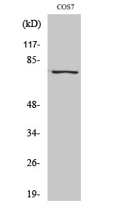
- Western Blot analysis of various cells using Phospho-NFκB-p65 (S529) Polyclonal Antibody diluted at 1:500

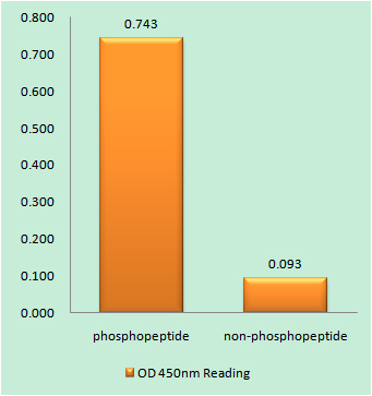
- Enzyme-Linked Immunosorbent Assay (Phospho-ELISA) for Immunogen Phosphopeptide (Phospho-left) and Non-Phosphopeptide (Phospho-right), using NF-κB p65 (Phospho-Ser529) Antibody

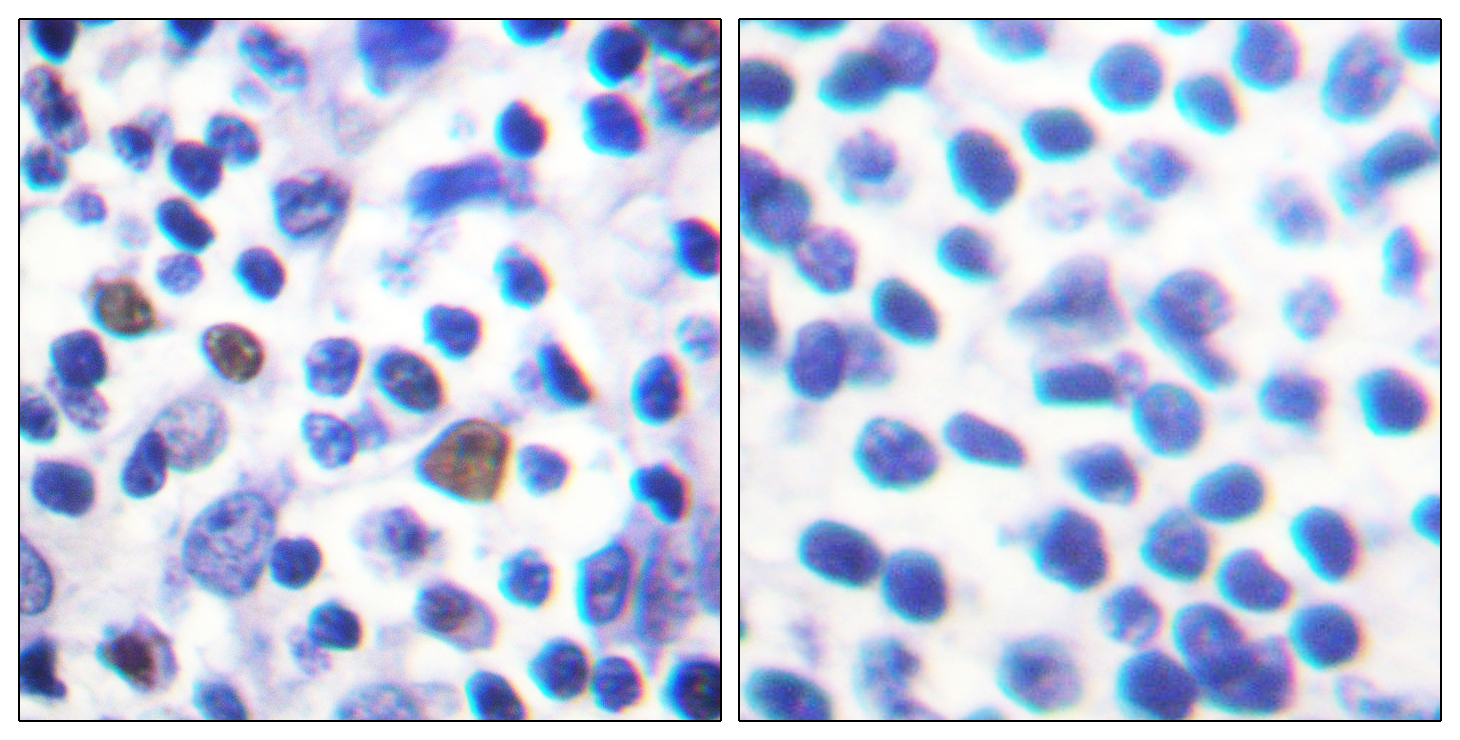
- Immunohistochemistry analysis of paraffin-embedded human breast cancer, using NF-κB p65 (Phospho-Ser529) Antibody. The picture on the right is blocked with the NF-κB p65 (Phospho-Ser529) peptide.


