Sp1 (phospho Thr739) Polyclonal Antibody
- Catalog No.:YP0248
- Applications:WB;IHC;IF;ELISA
- Reactivity:Human;Mouse;Rat;Monkey
- Target:
- Sp1
- Fields:
- >>Endocrine resistance;>>Mitophagy - animal;>>TGF-beta signaling pathway;>>Estrogen signaling pathway;>>Cortisol synthesis and secretion;>>Parathyroid hormone synthesis, secretion and action;>>Cushing syndrome;>>Huntington disease;>>Spinocerebellar ataxia;>>Human cytomegalovirus infection;>>Pathways in cancer;>>Transcriptional misregulation in cancer;>>Breast cancer;>>Choline metabolism in cancer;>>Diabetic cardiomyopathy
- Gene Name:
- SP1
- Protein Name:
- Transcription factor Sp1
- Human Gene Id:
- 6667
- Human Swiss Prot No:
- P08047
- Mouse Gene Id:
- 20683
- Mouse Swiss Prot No:
- O89090
- Rat Gene Id:
- 24790
- Rat Swiss Prot No:
- Q01714
- Immunogen:
- The antiserum was produced against synthesized peptide derived from human SP1 around the phosphorylation site of Thr739. AA range:706-755
- Specificity:
- Phospho-Sp1 (T739) Polyclonal Antibody detects endogenous levels of Sp1 protein only when phosphorylated at T739.
- Formulation:
- Liquid in PBS containing 50% glycerol, 0.5% BSA and 0.02% sodium azide.
- Source:
- Polyclonal, Rabbit,IgG
- Dilution:
- WB 1:500 - 1:2000. IHC 1:100 - 1:300. ELISA: 1:5000.. IF 1:50-200
- Purification:
- The antibody was affinity-purified from rabbit antiserum by affinity-chromatography using epitope-specific immunogen.
- Concentration:
- 1 mg/ml
- Storage Stability:
- -15°C to -25°C/1 year(Do not lower than -25°C)
- Other Name:
- SP1;TSFP1;Transcription factor Sp1
- Observed Band(KD):
- 81kD
- Background:
- The protein encoded by this gene is a zinc finger transcription factor that binds to GC-rich motifs of many promoters. The encoded protein is involved in many cellular processes, including cell differentiation, cell growth, apoptosis, immune responses, response to DNA damage, and chromatin remodeling. Post-translational modifications such as phosphorylation, acetylation, glycosylation, and proteolytic processing significantly affect the activity of this protein, which can be an activator or a repressor. Three transcript variants encoding different isoforms have been found for this gene. [provided by RefSeq, Nov 2014],
- Function:
- function:Binds to GC box promoters elements and selectively activates mRNA synthesis from genes that contain functional recognition sites. Can interact with G/C-rich motifs from serotonin receptor promoter.,PTM:O-glycosylated; contains N-acetylglucosamine side chains.,similarity:Belongs to the Sp1 C2H2-type zinc-finger protein family.,similarity:Contains 3 C2H2-type zinc fingers.,subunit:Interacts with ATF7IP, ATF7IP2, POGZ, HCFC1, AATF and PHC2. Interacts with varicella-zoster virus IE62 protein and HIV-1 Vpr. Interacts with SV40 VP2/3 proteins. Interacts with SV40 major capsid protein VP1; this interaction leads to a cooperativity between the two proteins in DNA binding.,
- Subcellular Location:
- Nucleus. Cytoplasm. Nuclear location is governed by glycosylated/phosphorylated states. Insulin promotes nuclear location, while glucagon favors cytoplasmic location.
- Expression:
- Up-regulated in adenocarcinomas of the stomach (at protein level). Isoform 3 is ubiquitously expressed at low levels.
- June 19-2018
- WESTERN IMMUNOBLOTTING PROTOCOL
- June 19-2018
- IMMUNOHISTOCHEMISTRY-PARAFFIN PROTOCOL
- June 19-2018
- IMMUNOFLUORESCENCE PROTOCOL
- September 08-2020
- FLOW-CYTOMEYRT-PROTOCOL
- May 20-2022
- Cell-Based ELISA│解您多样本WB检测之困扰
- July 13-2018
- CELL-BASED-ELISA-PROTOCOL-FOR-ACETYL-PROTEIN
- July 13-2018
- CELL-BASED-ELISA-PROTOCOL-FOR-PHOSPHO-PROTEIN
- July 13-2018
- Antibody-FAQs
- Products Images
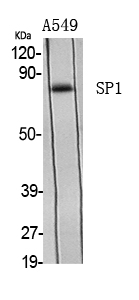
- Western Blot analysis of various cells using Phospho-Sp1 (T739) Polyclonal Antibody diluted at 1:1000
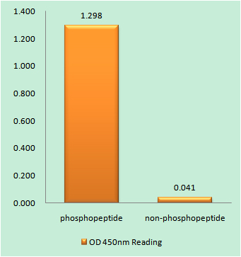
- Enzyme-Linked Immunosorbent Assay (Phospho-ELISA) for Immunogen Phosphopeptide (Phospho-left) and Non-Phosphopeptide (Phospho-right), using SP1 (Phospho-Thr739) Antibody
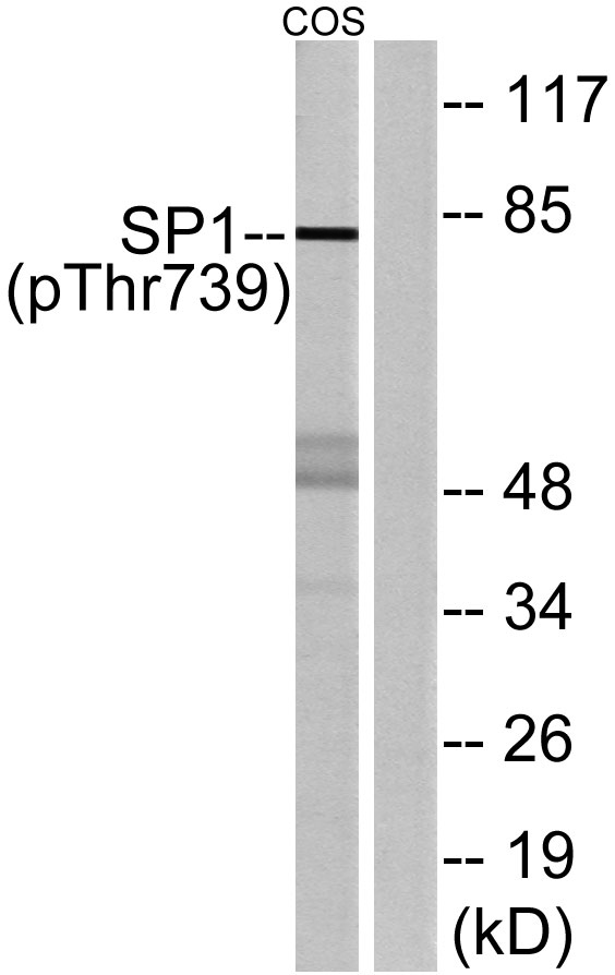
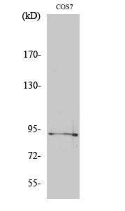
- Western blot analysis of lysates from COS7 cells treated with serum 20% 15', using SP1 (Phospho-Thr739) Antibody. The lane on the right is blocked with the phospho peptide.
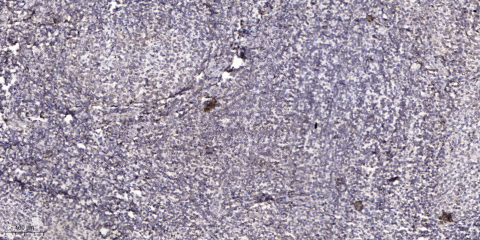
- Immunohistochemical analysis of paraffin-embedded human tonsil. 1, Antibody was diluted at 1:200(4° overnight). 2, Tris-EDTA,pH9.0 was used for antigen retrieval. 3,Secondary antibody was diluted at 1:200(room temperature, 45min).