Cyclin H (phospho Thr315) Polyclonal Antibody
- Catalog No.:YP0352
- Applications:WB;IHC;IF;ELISA
- Reactivity:Human;Mouse;Rat
- Target:
- Cyclin H
- Fields:
- >>Basal transcription factors;>>Nucleotide excision repair;>>Cell cycle
- Gene Name:
- CCNH
- Protein Name:
- Cyclin-H
- Human Gene Id:
- 902
- Human Swiss Prot No:
- P51946
- Mouse Gene Id:
- 66671
- Mouse Swiss Prot No:
- Q61458
- Rat Gene Id:
- 84389
- Rat Swiss Prot No:
- Q9R1A0
- Immunogen:
- The antiserum was produced against synthesized peptide derived from human Cyclin H around the phosphorylation site of Thr315. AA range:274-323
- Specificity:
- Phospho-Cyclin H (T315) Polyclonal Antibody detects endogenous levels of Cyclin H protein only when phosphorylated at T315.
- Formulation:
- Liquid in PBS containing 50% glycerol, 0.5% BSA and 0.02% sodium azide.
- Source:
- Polyclonal, Rabbit,IgG
- Dilution:
- WB 1:500 - 1:2000. IHC 1:100 - 1:300. ELISA: 1:10000.. IF 1:50-200
- Purification:
- The antibody was affinity-purified from rabbit antiserum by affinity-chromatography using epitope-specific immunogen.
- Concentration:
- 1 mg/ml
- Storage Stability:
- -15°C to -25°C/1 year(Do not lower than -25°C)
- Other Name:
- CCNH;Cyclin-H;MO15-associated protein;p34;p37
- Observed Band(KD):
- 34 36kD
- Background:
- The protein encoded by this gene belongs to the highly conserved cyclin family, whose members are characterized by a dramatic periodicity in protein abundance through the cell cycle. Cyclins function as regulators of CDK kinases. Different cyclins exhibit distinct expression and degradation patterns which contribute to the temporal coordination of each mitotic event. This cyclin forms a complex with CDK7 kinase and ring finger protein MAT1. The kinase complex is able to phosphorylate CDK2 and CDC2 kinases, thus functions as a CDK-activating kinase (CAK). This cyclin and its kinase partner are components of TFIIH, as well as RNA polymerase II protein complexes. They participate in two different transcriptional regulation processes, suggesting an important link between basal transcription control and the cell cycle machinery. A pseudogene of this gene is found on chromosome 4. Alternate splicing results in multiple t
- Function:
- function:Regulates CDK7, the catalytic subunit of the CDK-activating kinase (CAK) enzymatic complex. CAK activates the cyclin-associated kinases CDC2/CDK1, CDK2, CDK4 and CDK6 by threonine phosphorylation. CAK complexed to the core-TFIIH basal transcription factor activates RNA polymerase II by serine phosphorylation of the repetitive C-terminus domain (CTD) of its large subunit (POLR2A), allowing its escape from the promoter and elongation of the transcripts. Involved in cell cycle control and in RNA transcription by RNA polymerase II. Its expression and activity are constant throughout the cell cycle.,similarity:Belongs to the cyclin family.,similarity:Belongs to the cyclin family. Cyclin C subfamily.,subunit:Associates primarily with CDK7 and MAT1 to form the CAK complex. CAK can further associate with the core-TFIIH to form the TFIIH basal transcription factor.,
- Subcellular Location:
- Nucleus.
- Expression:
- Bone marrow,Brain,Embryonic brain,Epithelium,Liver,Urinary bladder,
- June 19-2018
- WESTERN IMMUNOBLOTTING PROTOCOL
- June 19-2018
- IMMUNOHISTOCHEMISTRY-PARAFFIN PROTOCOL
- June 19-2018
- IMMUNOFLUORESCENCE PROTOCOL
- September 08-2020
- FLOW-CYTOMEYRT-PROTOCOL
- May 20-2022
- Cell-Based ELISA│解您多样本WB检测之困扰
- July 13-2018
- CELL-BASED-ELISA-PROTOCOL-FOR-ACETYL-PROTEIN
- July 13-2018
- CELL-BASED-ELISA-PROTOCOL-FOR-PHOSPHO-PROTEIN
- July 13-2018
- Antibody-FAQs
- Products Images

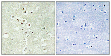
- Immunohistochemical analysis of paraffin-embedded Human brain. Antibody was diluted at 1:100(4° overnight). High-pressure and temperature Tris-EDTA,pH8.0 was used for antigen retrieval. Negetive contrl (right) obtaned from antibody was pre-absorbed by immunogen peptide.

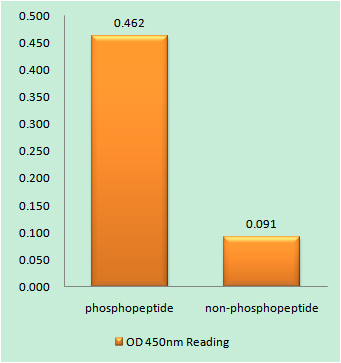
- Enzyme-Linked Immunosorbent Assay (Phospho-ELISA) for Immunogen Phosphopeptide (Phospho-left) and Non-Phosphopeptide (Phospho-right), using Cyclin H (Phospho-Thr315) Antibody

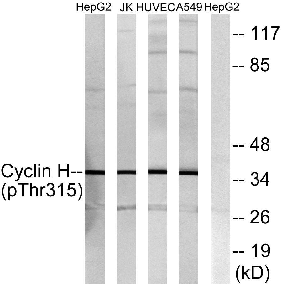
- Western blot analysis of lysates from HepG2 cells, Jurkat cells, HUVEC cells and A549 cells, using Cyclin H (Phospho-Thr315) Antibody. The lane on the right is blocked with the phospho peptide.


