DAPK3 (phospho Thr265) Polyclonal Antibody
- Catalog No.:YP0548
- Applications:WB;IHC;IF;ELISA
- Reactivity:Human;Mouse;Rat
- Target:
- DAPK3
- Fields:
- >>Autophagy - animal;>>Pathways in cancer;>>Bladder cancer
- Gene Name:
- DAPK3
- Protein Name:
- Death-associated protein kinase 3
- Human Gene Id:
- 1613
- Human Swiss Prot No:
- O43293
- Mouse Gene Id:
- 13144
- Mouse Swiss Prot No:
- O54784
- Rat Gene Id:
- 64391
- Rat Swiss Prot No:
- O88764
- Immunogen:
- The antiserum was produced against synthesized peptide derived from human DAPK3 around the phosphorylation site of Thr265. AA range:241-290
- Specificity:
- Phospho-DAPK3 (T265) Polyclonal Antibody detects endogenous levels of DAPK3 protein only when phosphorylated at T265.
- Formulation:
- Liquid in PBS containing 50% glycerol, 0.5% BSA and 0.02% sodium azide.
- Source:
- Polyclonal, Rabbit,IgG
- Dilution:
- WB 1:500 - 1:2000. IHC 1:100 - 1:300. IF 1:200 - 1:1000. ELISA: 1:10000. Not yet tested in other applications.
- Purification:
- The antibody was affinity-purified from rabbit antiserum by affinity-chromatography using epitope-specific immunogen.
- Concentration:
- 1 mg/ml
- Storage Stability:
- -15°C to -25°C/1 year(Do not lower than -25°C)
- Other Name:
- DAPK3;ZIPK;Death-associated protein kinase 3;DAP kinase 3;DAP-like kinase;Dlk;MYPT1 kinase;Zipper-interacting protein kinase;ZIP-kinase
- Observed Band(KD):
- 52kD
- Background:
- Death-associated protein kinase 3 (DAPK3) induces morphological changes in apoptosis when overexpressed in mammalian cells. These results suggest that DAPK3 may play a role in the induction of apoptosis. [provided by RefSeq, Jul 2008],
- Function:
- catalytic activity:ATP + a protein = ADP + a phosphoprotein.,cofactor:Magnesium.,function:Serine/threonine kinase which acts as a positive regulator of apoptosis. Phosphorylates histone H3 on 'Thr-11' at centromeres during mitosis.,similarity:Belongs to the protein kinase superfamily. CAMK Ser/Thr protein kinase family. DAP kinase subfamily.,similarity:Contains 1 protein kinase domain.,subcellular location:Relocates to the cytoplasm on binding PAWR where the complex appears to interact with actin filaments (By similarity). Associates to centromeres from prophase to anaphase.,subunit:Homodimer or forms heterodimers with ATF4. Both interactions require an intact leucine zipper domain and oligomerization is required for full enzymatic activity. Also binds to DAXX and PAWR, possibly in a ternary complex which plays a role in caspase activation. Interacts with AATF and CDC5L.,
- Subcellular Location:
- Nucleus . Cytoplasm . Predominantly localizes to the cytoplasm but can shuttle between the nucleus and cytoplasm; cytoplasmic localization is promoted by phosphorylation at Thr-299 and involves Rho/Rock signaling. .; [Isoform 1]: Nucleus . Cytoplasm .; [Isoform 2]: Nucleus . Cytoplasm .
- Expression:
- Widely expressed. Isoform 1 and isoform 2 are expressed in the bladder smooth muscle.
- June 19-2018
- WESTERN IMMUNOBLOTTING PROTOCOL
- June 19-2018
- IMMUNOHISTOCHEMISTRY-PARAFFIN PROTOCOL
- June 19-2018
- IMMUNOFLUORESCENCE PROTOCOL
- September 08-2020
- FLOW-CYTOMEYRT-PROTOCOL
- May 20-2022
- Cell-Based ELISA│解您多样本WB检测之困扰
- July 13-2018
- CELL-BASED-ELISA-PROTOCOL-FOR-ACETYL-PROTEIN
- July 13-2018
- CELL-BASED-ELISA-PROTOCOL-FOR-PHOSPHO-PROTEIN
- July 13-2018
- Antibody-FAQs
- Products Images
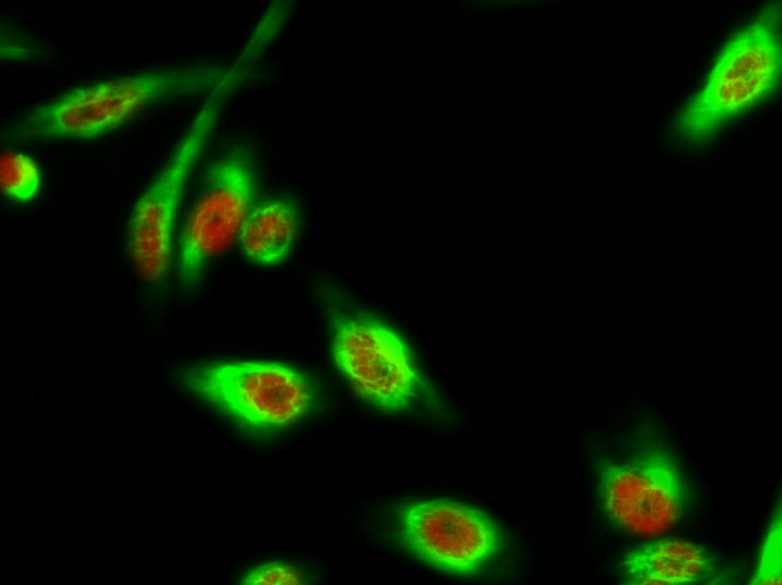
- Immunofluorescence analysis of Hela cell. 1,DAPK3 (phospho Thr265) Polyclonal Antibody(red) was diluted at 1:200(4° overnight). α-tubulin Monoclonal Antibody(8F11)(green) was diluted at 1:200(4° overnight). 2, Goat Anti Rabbit Alexa Fluor 594 Catalog:RS3611 was diluted at 1:1000(room temperature, 50min). Goat Anti Mouse Alexa Fluor 488 Catalog:RS3208 was diluted at 1:1000(room temperature, 50min).
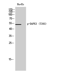
- Western Blot analysis of HuvEc cells using Phospho-DAPK3 (T265) Polyclonal Antibody
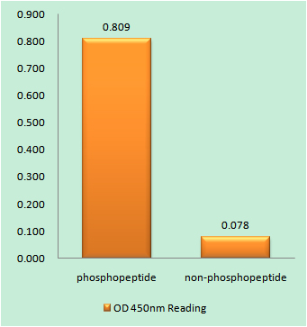
- Enzyme-Linked Immunosorbent Assay (Phospho-ELISA) for Immunogen Phosphopeptide (Phospho-left) and Non-Phosphopeptide (Phospho-right), using DAPK3 (Phospho-Thr265) Antibody

- Immunofluorescence analysis of A549 cells, using DAPK3 (Phospho-Thr265) Antibody. The picture on the right is blocked with the phospho peptide.

- Western blot analysis of lysates from HUVEC cells, using DAPK3 (Phospho-Thr265) Antibody. The lane on the right is blocked with the phospho peptide.

- Immunohistochemical analysis of paraffin-embedded human tonsil. 1, Antibody was diluted at 1:200(4° overnight). 2, Tris-EDTA,pH9.0 was used for antigen retrieval. 3,Secondary antibody was diluted at 1:200(room temperature, 45min).


