Lck (phospho Tyr393) Polyclonal Antibody
- Catalog No.:YP0572
- Applications:WB;IF;ELISA
- Reactivity:Human;Mouse;Rat
- Target:
- Lck
- Fields:
- >>NF-kappa B signaling pathway;>>Osteoclast differentiation;>>Natural killer cell mediated cytotoxicity;>>Th1 and Th2 cell differentiation;>>Th17 cell differentiation;>>T cell receptor signaling pathway;>>Yersinia infection;>>Human T-cell leukemia virus 1 infection;>>PD-L1 expression and PD-1 checkpoint pathway in cancer;>>Primary immunodeficiency
- Gene Name:
- LCK
- Protein Name:
- Tyrosine-protein kinase Lck
- Human Gene Id:
- 3932
- Human Swiss Prot No:
- P06239
- Mouse Gene Id:
- 16818
- Mouse Swiss Prot No:
- P06240
- Rat Gene Id:
- 313050
- Rat Swiss Prot No:
- Q01621
- Immunogen:
- The antiserum was produced against synthesized peptide derived from human Lck around the phosphorylation site of Tyr393. AA range:361-410
- Specificity:
- Phospho-Lck (Y393) Polyclonal Antibody detects endogenous levels of Lck protein only when phosphorylated at Y393.
- Formulation:
- Liquid in PBS containing 50% glycerol, 0.5% BSA and 0.02% sodium azide.
- Source:
- Polyclonal, Rabbit,IgG
- Dilution:
- WB 1:500 - 1:2000. IF 1:200 - 1:1000. ELISA: 1:10000. Not yet tested in other applications.
- Purification:
- The antibody was affinity-purified from rabbit antiserum by affinity-chromatography using epitope-specific immunogen.
- Concentration:
- 1 mg/ml
- Storage Stability:
- -15°C to -25°C/1 year(Do not lower than -25°C)
- Other Name:
- LCK;Tyrosine-protein kinase Lck;Leukocyte C-terminal Src kinase;LSK;Lymphocyte cell-specific protein-tyrosine kinase;Protein YT16;Proto-oncogene Lck;T cell-specific protein-tyrosine kinase;p56-LCK
- Observed Band(KD):
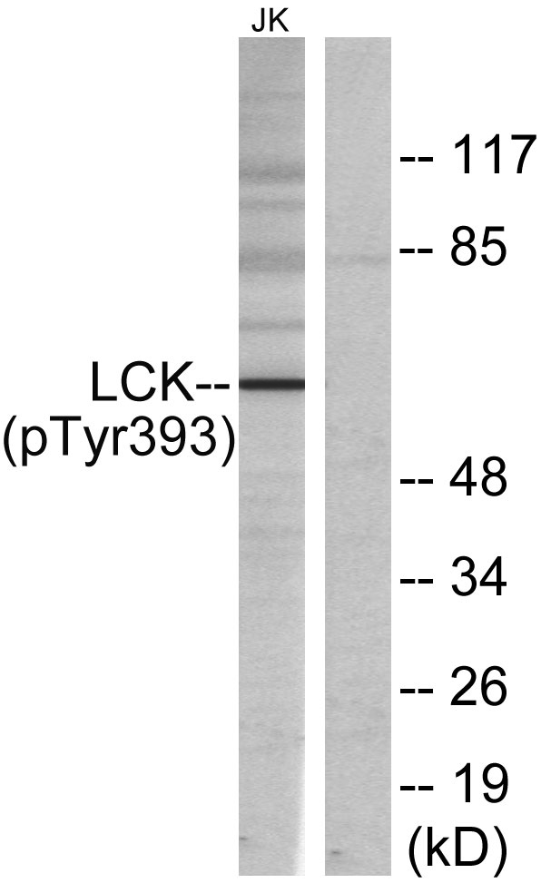
- 60kD
- Background:
- This gene is a member of the Src family of protein tyrosine kinases (PTKs). The encoded protein is a key signaling molecule in the selection and maturation of developing T-cells. It contains N-terminal sites for myristylation and palmitylation, a PTK domain, and SH2 and SH3 domains which are involved in mediating protein-protein interactions with phosphotyrosine-containing and proline-rich motifs, respectively. The protein localizes to the plasma membrane and pericentrosomal vesicles, and binds to cell surface receptors, including CD4 and CD8, and other signaling molecules. Multiple alternatively spliced variants encoding different isoforms have been described. [provided by RefSeq, Aug 2016],
- Function:
- catalytic activity:ATP + a [protein]-L-tyrosine = ADP + a [protein]-L-tyrosine phosphate.,disease:A chromosomal aberration involving LCK is found in leukemias. Translocation t(1;7)(p34;q34) with TCRB.,domain:The SH2 domain mediates interaction with SQSTM1. Interaction is regulated by Ser-59 phosphorylation.,enzyme regulation:Inhibited by tyrosine phosphorylation.,function:Tyrosine kinase that plays an essential role for the selection and maturation of developing T-cell in the thymus and in mature T-cell function. Is constitutively associated with the cytoplasmic portions of the CD4 and CD8 surface receptors and plays a key role in T-cell antigen receptor(TCR)-linked signal transduction pathways. Association of the TCR with a peptide antigen-bound MHC complex facilitates the interaction of CD4 and CD8 with MHC class II and class I molecules, respectively, and thereby recruits the associat
- Subcellular Location:
- Cell membrane ; Lipid-anchor ; Cytoplasmic side . Cytoplasm, cytosol . Present in lipid rafts in an inactive form. .
- Expression:
- Expressed specifically in lymphoid cells.
- June 19-2018
- WESTERN IMMUNOBLOTTING PROTOCOL
- June 19-2018
- IMMUNOHISTOCHEMISTRY-PARAFFIN PROTOCOL
- June 19-2018
- IMMUNOFLUORESCENCE PROTOCOL
- September 08-2020
- FLOW-CYTOMEYRT-PROTOCOL
- May 20-2022
- Cell-Based ELISA│解您多样本WB检测之困扰
- July 13-2018
- CELL-BASED-ELISA-PROTOCOL-FOR-ACETYL-PROTEIN
- July 13-2018
- CELL-BASED-ELISA-PROTOCOL-FOR-PHOSPHO-PROTEIN
- July 13-2018
- Antibody-FAQs
- Products Images

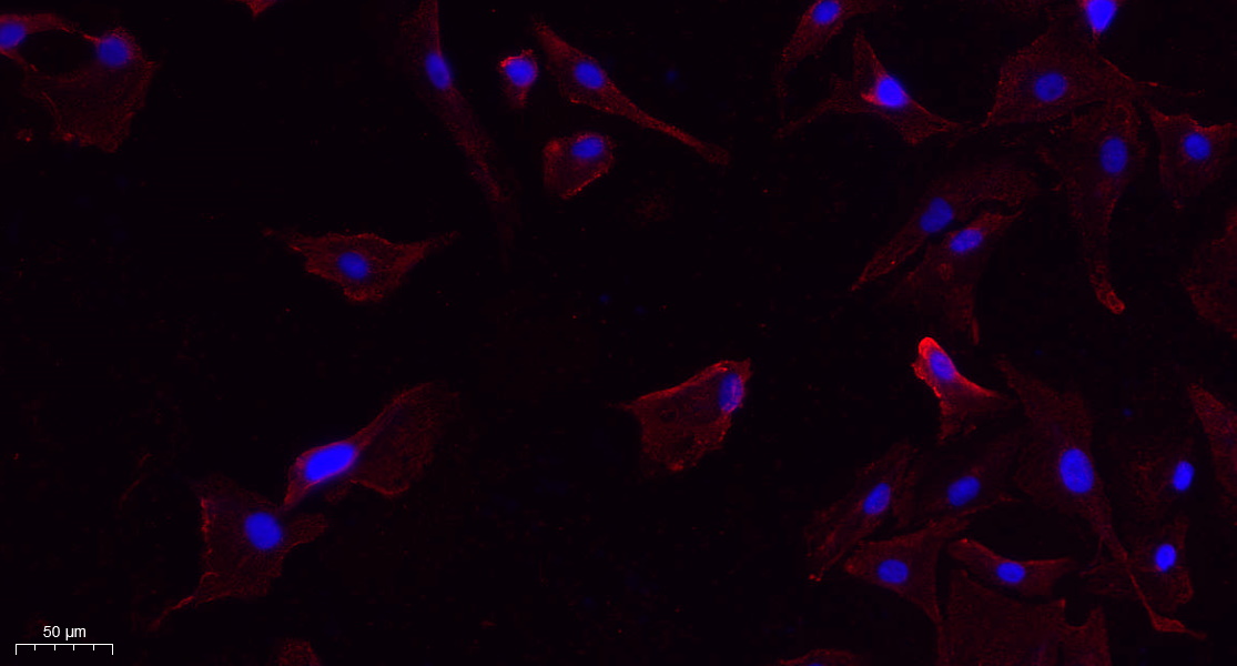
- Immunofluorescence analysis of A549. 1,primary Antibody(red) was diluted at 1:200(4°C overnight). 2, Goat Anti Rabbit IgG (H&L) - Alexa Fluor 594 Secondary antibody was diluted at 1:1000(room temperature, 50min).3, Picture B: DAPI(blue) 10min.

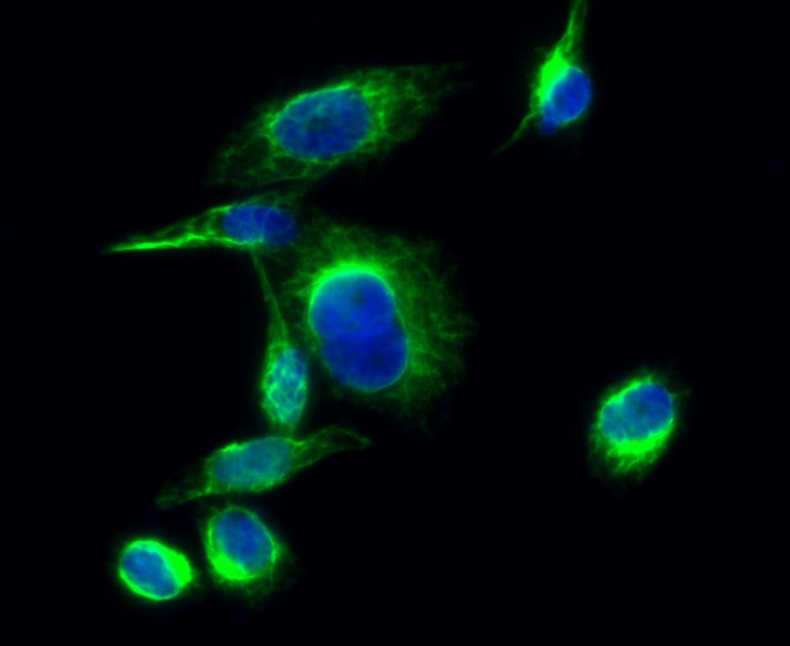
- Immunofluorescence analysis of Hela cell. 1,Lck (phospho Tyr393) Polyclonal Antibody(green) was diluted at 1:200(4° overnight). 2, Goat Anti Rabbit Alexa Fluor 488 Catalog:RS3211 was diluted at 1:1000(room temperature, 50min). 3 DAPI(blue) 10min.

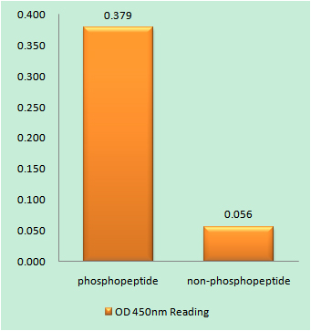
- Enzyme-Linked Immunosorbent Assay (Phospho-ELISA) for Immunogen Phosphopeptide (Phospho-left) and Non-Phosphopeptide (Phospho-right), using Lck (Phospho-Tyr393) Antibody

- Western blot analysis of lysates from Jurkat cells, using Lck (Phospho-Tyr393) Antibody. The lane on the right is blocked with the phospho peptide.


