Ah Receptor (phospho Ser36) Polyclonal Antibody
- Catalog No.:YP0713
- Applications:WB;IHC;IF;ELISA
- Reactivity:Human;Mouse;Rat
- Target:
- Ah Receptor
- Fields:
- >>Th17 cell differentiation;>>Cushing syndrome;>>Chemical carcinogenesis - receptor activation;>>Chemical carcinogenesis - reactive oxygen species
- Gene Name:
- AHR
- Protein Name:
- Aryl hydrocarbon receptor
- Human Gene Id:
- 196/57491
- Human Swiss Prot No:
- P35869/A9YTQ3
- Mouse Gene Id:
- 11622/11624
- Rat Gene Id:
- 25690/498999
- Rat Swiss Prot No:
- P41738/Q75NT5
- Immunogen:
- The antiserum was produced against synthesized peptide derived from human AhR around the phosphorylation site of Ser36. AA range:2-51
- Specificity:
- Phospho-Ah Receptor (S36) Polyclonal Antibody detects endogenous levels of Ah Receptor protein only when phosphorylated at S36.
- Formulation:
- Liquid in PBS containing 50% glycerol, 0.5% BSA and 0.02% sodium azide.
- Source:
- Polyclonal, Rabbit,IgG
- Dilution:
- WB 1:500 - 1:2000. IHC 1:100 - 1:300. ELISA: 1:5000.. IF 1:50-200
- Purification:
- The antibody was affinity-purified from rabbit antiserum by affinity-chromatography using epitope-specific immunogen.
- Concentration:
- 1 mg/ml
- Storage Stability:
- -15°C to -25°C/1 year(Do not lower than -25°C)
- Other Name:
- AHR;BHLHE76;Aryl hydrocarbon receptor;Ah receptor;AhR;Class E basic helix-loop-helix protein 76;bHLHe76;AHRR;BHLHE77;KIAA1234;Aryl hydrocarbon receptor repressor;AhR repressor;AhRR;Class E basic helix-loop-helix protein 77;bHL
- Observed Band(KD):
- 75 or 96kD
- Background:
- The protein encoded by this gene is a ligand-activated helix-loop-helix transcription factor involved in the regulation of biological responses to planar aromatic hydrocarbons. This receptor has been shown to regulate xenobiotic-metabolizing enzymes such as cytochrome P450. Before ligand binding, the encoded protein is sequestered in the cytoplasm; upon ligand binding, this protein moves to the nucleus and stimulates transcription of target genes. [provided by RefSeq, Sep 2015],
- Function:
- function:Ligand-activated transcriptional activator. Binds to the XRE promoter region of genes it activates. Activates the expression of multiple phase I and II xenobiotic chemical metabolizing enzyme genes (such as the CYP1A1 gene). Mediates biochemical and toxic effects of halogenated aromatic hydrocarbons. Involved in cell-cycle regulation. Likely to play an important role in the development and maturation of many tissues.,induction:Induced or repressed by TGF-beta and dioxin in a cell-type specific fashion. Repressed by cAMP, retinoic acid, and TPA.,similarity:Contains 1 basic helix-loop-helix (bHLH) domain.,similarity:Contains 1 PAC (PAS-associated C-terminal) domain.,similarity:Contains 2 PAS (PER-ARNT-SIM) domains.,subcellular location:Initially cytoplasmic; upon binding with ligand and interaction with a HSP90, it translocates to the nucleus.,subunit:Binds MYBBP1A (By similarity)
- Subcellular Location:
- Cytoplasm . Nucleus . Initially cytoplasmic; upon binding with ligand and interaction with a HSP90, it translocates to the nucleus. .
- Expression:
- Expressed in all tissues tested including blood, brain, heart, kidney, liver, lung, pancreas and skeletal muscle. Expressed in retinal photoreceptors (PubMed:29726989).
- June 19-2018
- WESTERN IMMUNOBLOTTING PROTOCOL
- June 19-2018
- IMMUNOHISTOCHEMISTRY-PARAFFIN PROTOCOL
- June 19-2018
- IMMUNOFLUORESCENCE PROTOCOL
- September 08-2020
- FLOW-CYTOMEYRT-PROTOCOL
- May 20-2022
- Cell-Based ELISA│解您多样本WB检测之困扰
- July 13-2018
- CELL-BASED-ELISA-PROTOCOL-FOR-ACETYL-PROTEIN
- July 13-2018
- CELL-BASED-ELISA-PROTOCOL-FOR-PHOSPHO-PROTEIN
- July 13-2018
- Antibody-FAQs
- Products Images

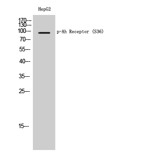
- Western Blot analysis of HepG2 cells using Phospho-Ah Receptor (S36) Polyclonal Antibody

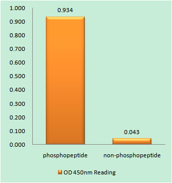
- Enzyme-Linked Immunosorbent Assay (Phospho-ELISA) for Immunogen Phosphopeptide (Phospho-left) and Non-Phosphopeptide (Phospho-right), using AhR (Phospho-Ser36) Antibody

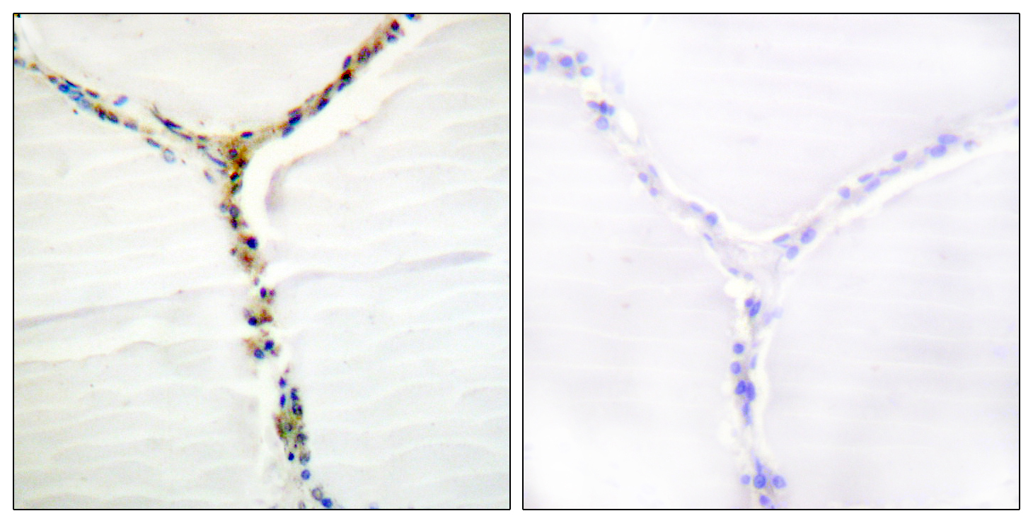
- Immunohistochemistry analysis of paraffin-embedded human thyroid gland, using AhR (Phospho-Ser36) Antibody. The picture on the right is blocked with the phospho peptide.

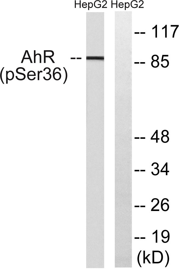
- Western blot analysis of lysates from HepG2 cells, using AhR (Phospho-Ser36) Antibody. The lane on the right is blocked with the phospho peptide.


