MRLC2 (phospho Ser20) Polyclonal Antibody
- Catalog No.:YP0901
- Applications:WB;IHC;IF;ELISA
- Reactivity:Human;Mouse;Rat
- Target:
- Myosin Light Chain 2
- Fields:
- >>cGMP-PKG signaling pathway;>>cAMP signaling pathway;>>Vascular smooth muscle contraction;>>Axon guidance;>>Focal adhesion;>>Tight junction;>>Leukocyte transendothelial migration;>>Regulation of actin cytoskeleton;>>Oxytocin signaling pathway;>>Shigellosis;>>Salmonella infection
- Gene Name:
- MYL9
- Protein Name:
- Myosin regulatory light polypeptide 9
- Human Gene Id:
- 10398/10627
- Human Swiss Prot No:
- P24844
- Mouse Gene Id:
- 98932
- Mouse Swiss Prot No:
- Q9CQ19
- Rat Gene Id:
- 296313
- Rat Swiss Prot No:
- Q64122
- Immunogen:
- The antiserum was produced against synthesized peptide derived from human Myosin regulatory light chain 2 around the phosphorylation site of Ser18. AA range:3-52
- Specificity:
- Phospho-MRLC2 (S20) Polyclonal Antibody detects endogenous levels of MRLC2 protein only when phosphorylated at S20.
- Formulation:
- Liquid in PBS containing 50% glycerol, 0.5% BSA and 0.02% sodium azide.
- Source:
- Polyclonal, Rabbit,IgG
- Dilution:
- WB 1:500 - 1:2000. IHC 1:100 - 1:300. IF 1:200 - 1:1000. ELISA: 1:20000. Not yet tested in other applications.
- Purification:
- The antibody was affinity-purified from rabbit antiserum by affinity-chromatography using epitope-specific immunogen.
- Concentration:
- 1 mg/ml
- Storage Stability:
- -15°C to -25°C/1 year(Do not lower than -25°C)
- Other Name:
- MYL9;MLC2;MRLC1;MYRL2;Myosin regulatory light polypeptide 9;20 kDa myosin light chain;LC20;MLC-2C;Myosin RLC;Myosin regulatory light chain 2; smooth muscle isoform;Myosin regulatory light chain 9;Myosin regulatory light chain MRL
- Observed Band(KD):
- 18kD
- Background:
- Myosin, a structural component of muscle, consists of two heavy chains and four light chains. The protein encoded by this gene is a myosin light chain that may regulate muscle contraction by modulating the ATPase activity of myosin heads. The encoded protein binds calcium and is activated by myosin light chain kinase. Two transcript variants encoding different isoforms have been found for this gene. [provided by RefSeq, Jul 2008],
- Function:
- function:Myosin regulatory subunit that plays an important role in regulation of both smooth muscle and nonmuscle cell contractile activity via its phosphorylation. Implicated in cytokinesis, receptor capping, and cell locomotion.,miscellaneous:This chain binds calcium.,PTM:Phosphorylation increases the actin-activated myosin ATPase activity and thereby regulates the contractile activity. It is required to generate the driving force in the migration of the cells but not necessary for localization of myosin-2 at the leading edge.,similarity:Contains 3 EF-hand domains.,subunit:Myosin is an hexamer of 2 heavy chains and 4 light chains.,tissue specificity:Smooth muscle tissues and in some, but not all, nonmuscle cells.,
- Subcellular Location:
- Cytoplasm, cytoskeleton . Cytoplasm, cell cortex . Colocalizes with F-actin, MYH9 and PIEZO1 at the actomyosin cortex in myoblasts. .
- Expression:
- Smooth muscle tissues and in some, but not all, nonmuscle cells.
- June 19-2018
- WESTERN IMMUNOBLOTTING PROTOCOL
- June 19-2018
- IMMUNOHISTOCHEMISTRY-PARAFFIN PROTOCOL
- June 19-2018
- IMMUNOFLUORESCENCE PROTOCOL
- September 08-2020
- FLOW-CYTOMEYRT-PROTOCOL
- May 20-2022
- Cell-Based ELISA│解您多样本WB检测之困扰
- July 13-2018
- CELL-BASED-ELISA-PROTOCOL-FOR-ACETYL-PROTEIN
- July 13-2018
- CELL-BASED-ELISA-PROTOCOL-FOR-PHOSPHO-PROTEIN
- July 13-2018
- Antibody-FAQs
- Products Images
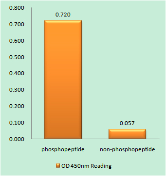
- Enzyme-Linked Immunosorbent Assay (Phospho-ELISA) for Immunogen Phosphopeptide (Phospho-left) and Non-Phosphopeptide (Phospho-right), using Myosin regulatory light chain 2 (Phospho-Ser18) Antibody
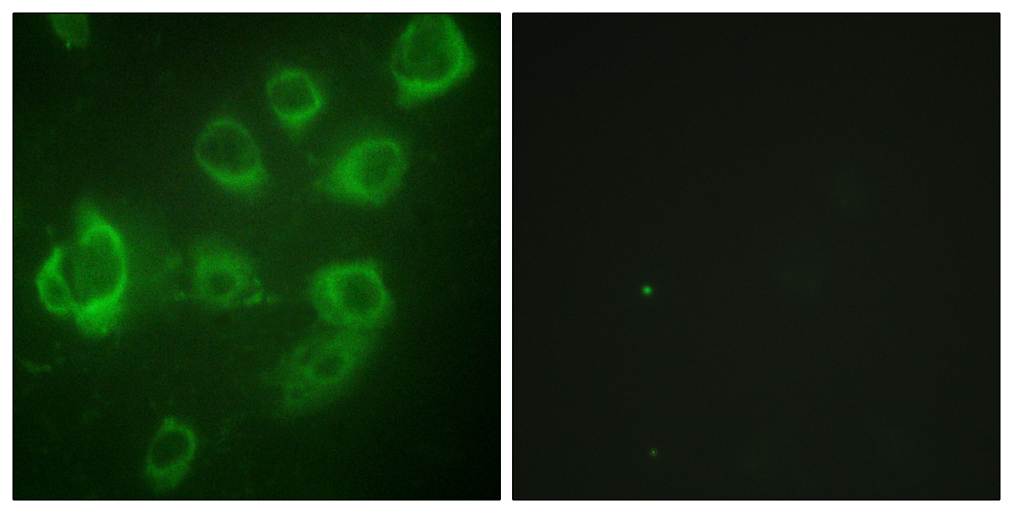
- Immunofluorescence analysis of HUVEC cells, using Myosin regulatory light chain 2 (Phospho-Ser18) Antibody. The picture on the right is blocked with the phospho peptide.
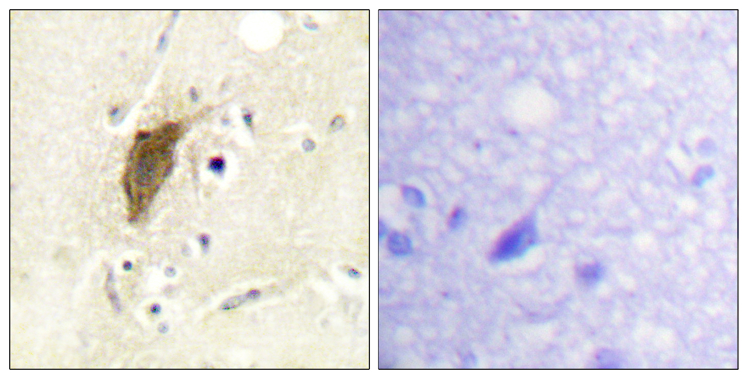
- Immunohistochemistry analysis of paraffin-embedded human brain, using Myosin regulatory light chain 2 (Phospho-Ser18) Antibody. The picture on the right is blocked with the phospho peptide.

- Western blot analysis of lysates from LOVO cells treated with H2O2 100uM 30', using Myosin regulatory light chain 2 (Phospho-Ser18) Antibody. The lane on the right is blocked with the phospho peptide.
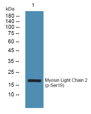
- Western blot analysis of lysates from PC12 cells, primary antibody was diluted at 1:1000, 4°over night


