PAK4 (phospho Ser474) Polyclonal Antibody
- Catalog No.:YP0973
- Applications:WB;IHC;IF;ELISA
- Reactivity:Human;Mouse;Rat
- Target:
- PAK4
- Fields:
- >>ErbB signaling pathway;>>Ras signaling pathway;>>Axon guidance;>>Focal adhesion;>>T cell receptor signaling pathway;>>Regulation of actin cytoskeleton;>>Human immunodeficiency virus 1 infection;>>MicroRNAs in cancer;>>Renal cell carcinoma
- Gene Name:
- PAK4
- Protein Name:
- Serine/threonine-protein kinase PAK 4
- Human Gene Id:
- 10298
- Human Swiss Prot No:
- O96013
- Mouse Gene Id:
- 70584
- Mouse Swiss Prot No:
- Q8BTW9
- Immunogen:
- The antiserum was produced against synthesized peptide derived from human PAK4/5/6 around the phosphorylation site of Ser474. AA range:441-490
- Specificity:
- Phospho-PAK4 (S474) Polyclonal Antibody detects endogenous levels of PAK4 protein only when phosphorylated at S474.
- Formulation:
- Liquid in PBS containing 50% glycerol, 0.5% BSA and 0.02% sodium azide.
- Source:
- Polyclonal, Rabbit,IgG
- Dilution:
- WB 1:500 - 1:2000. IHC 1:100 - 1:300. ELISA: 1:20000.. IF 1:50-200
- Purification:
- The antibody was affinity-purified from rabbit antiserum by affinity-chromatography using epitope-specific immunogen.
- Concentration:
- 1 mg/ml
- Storage Stability:
- -15°C to -25°C/1 year(Do not lower than -25°C)
- Other Name:
- PAK4;KIAA1142;Serine/threonine-protein kinase PAK 4;p21-activated kinase 4;PAK-4
- Observed Band(KD):
- 64kD
- Background:
- PAK proteins, a family of serine/threonine p21-activating kinases, include PAK1, PAK2, PAK3 and PAK4. PAK proteins are critical effectors that link Rho GTPases to cytoskeleton reorganization and nuclear signaling. They serve as targets for the small GTP binding proteins Cdc42 and Rac and have been implicated in a wide range of biological activities. PAK4 interacts specifically with the GTP-bound form of Cdc42Hs and weakly activates the JNK family of MAP kinases. PAK4 is a mediator of filopodia formation and may play a role in the reorganization of the actin cytoskeleton. Multiple alternatively spliced transcript variants encoding distinct isoforms have been found for this gene. [provided by RefSeq, Jul 2008],
- Function:
- catalytic activity:ATP + a protein = ADP + a phosphoprotein.,function:Activates the JNK pathway. Plays a role in the reorganization of the actin cytoskeleton and in the formation of filopodia. Phosphorylates and inactivates the protein phosphatase SSH1, leading to increased inhibitory phosphorylation of the actin binding/depolymerizing factor cofilin. Decreased cofilin activity may lead to stabilization of actin filaments. Phosphorylates ARHGEF2.,PTM:Autophosphorylated on serine residues when activated by CDC42/p21.,PTM:Phosphorylated on tyrosine residues upon stimulation of FGFR2.,similarity:Belongs to the protein kinase superfamily. STE Ser/Thr protein kinase family. STE20 subfamily.,similarity:Contains 1 CRIB domain.,similarity:Contains 1 protein kinase domain.,subunit:Interacts with FGFR2 and GRB2 (By similarity). Interacts tightly with GTP-bound but not GDP-bound CDC42/p21 and weakl
- Subcellular Location:
- Cytoplasm . Seems to shuttle between cytoplasmic compartments depending on the activating effector. For example, can be found on the cell periphery after activation of growth-factor or integrin-mediated signaling pathways. .
- Expression:
- Highest expression in prostate, testis and colon.
- June 19-2018
- WESTERN IMMUNOBLOTTING PROTOCOL
- June 19-2018
- IMMUNOHISTOCHEMISTRY-PARAFFIN PROTOCOL
- June 19-2018
- IMMUNOFLUORESCENCE PROTOCOL
- September 08-2020
- FLOW-CYTOMEYRT-PROTOCOL
- May 20-2022
- Cell-Based ELISA│解您多样本WB检测之困扰
- July 13-2018
- CELL-BASED-ELISA-PROTOCOL-FOR-ACETYL-PROTEIN
- July 13-2018
- CELL-BASED-ELISA-PROTOCOL-FOR-PHOSPHO-PROTEIN
- July 13-2018
- Antibody-FAQs
- Products Images

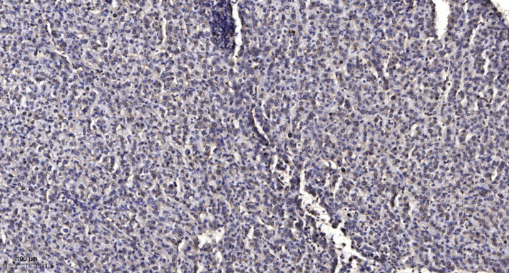
- Immunohistochemical analysis of paraffin-embedded human liver cancer. 1, Antibody was diluted at 1:200(4° overnight). 2, Tris-EDTA,pH9.0 was used for antigen retrieval. 3,Secondary antibody was diluted at 1:200(room temperature, 45min).

- Western Blot analysis of various,using primary antibody at 1:1000 dilution. Secondary antibody(catalog#:RS23920) was diluted at 1:10000


