Rev-erbα (Phospho Ser55/59) rabbit pAb
- Catalog No.:YP1465
- Applications:WB;IHC
- Reactivity:Human;Rat;Mouse;
- Target:
- Rev-erbα
- Fields:
- >>Circadian rhythm
- Gene Name:
- NR1D1 EAR1 HREV THRAL
- Protein Name:
- Rev-erbα (Ser55/59)
- Human Gene Id:
- 9572
- Human Swiss Prot No:
- P20393
- Mouse Gene Id:
- 217166
- Mouse Swiss Prot No:
- Q3UV55
- Rat Gene Id:
- 252917
- Rat Swiss Prot No:
- Q63503
- Immunogen:
- Synthesized phosho peptide around human Rev-erbα (Ser55 and 59)
- Specificity:
- This antibody detects endogenous levels of Human Rev-erbα (phospho-Ser55 or 59)
- Formulation:
- Liquid in PBS containing 50% glycerol, 0.5% BSA and 0.02% sodium azide.
- Source:
- Polyclonal, Rabbit,IgG
- Dilution:
- WB 1:500-2000;IHC 1:50-300
- Purification:
- The antibody was affinity-purified from rabbit serum by affinity-chromatography using specific immunogen.
- Concentration:
- 1 mg/ml
- Storage Stability:
- -15°C to -25°C/1 year(Do not lower than -25°C)
- Other Name:
- Nuclear receptor subfamily 1 group D member 1 (Rev-erbA-alpha) (V-erbA-related protein 1) (EAR-1)
- Observed Band(KD):
- 67kD
- Background:
- This gene encodes a transcription factor that is a member of the nuclear receptor subfamily 1. The encoded protein is a ligand-sensitive transcription factor that negatively regulates the expression of core clock proteins. In particular this protein represses the circadian clock transcription factor aryl hydrocarbon receptor nuclear translocator-like protein 1 (ARNTL). This protein may also be involved in regulating genes that function in metabolic, inflammatory and cardiovascular processes. [provided by RefSeq, Jan 2013],
- Function:
- domain:Composed of three domains: a modulating N-terminal domain, a DNA-binding domain and a C-terminal steroid-binding domain.,function:Functions as a constitutive transcriptional repressor. Possible receptor for triiodothyronine.,similarity:Belongs to the nuclear hormone receptor family. NR1 subfamily.,similarity:Contains 1 nuclear receptor DNA-binding domain.,subunit:Interacts with C1D.,tissue specificity:Expressed in all tissues and cell lines examined. Expressed at high levels in some squamous carcinoma cell lines.,
- Subcellular Location:
- Nucleus . Cytoplasm . Cell projection, dendrite . Cell projection, dendritic spine . Localizes to the cytoplasm, dendrites and dendritic spine in the presence of OPHN1. Localizes predominantly to the nucleus at ZT8 whereas it is cytoplasmic at ZT20. Phosphorylation by CSNK1E enhances its cytoplasmic localization. .
- Expression:
- Widely expressed. Expressed at high levels in the liver, adipose tissue, skeletal muscle and brain. Also expressed in endothelial cells (ECs), vascular smooth muscle cells (VSMCs) and macrophages. Expression oscillates diurnally in the suprachiasmatic nucleus (SCN) of the hypothalamus as well as in peripheral tissues. Expression increases during the differentiation of pre-adipocytes into mature adipocytes. Expressed at high levels in some squamous carcinoma cell lines.
- June 19-2018
- WESTERN IMMUNOBLOTTING PROTOCOL
- June 19-2018
- IMMUNOHISTOCHEMISTRY-PARAFFIN PROTOCOL
- June 19-2018
- IMMUNOFLUORESCENCE PROTOCOL
- September 08-2020
- FLOW-CYTOMEYRT-PROTOCOL
- May 20-2022
- Cell-Based ELISA│解您多样本WB检测之困扰
- July 13-2018
- CELL-BASED-ELISA-PROTOCOL-FOR-ACETYL-PROTEIN
- July 13-2018
- CELL-BASED-ELISA-PROTOCOL-FOR-PHOSPHO-PROTEIN
- July 13-2018
- Antibody-FAQs
- Products Images

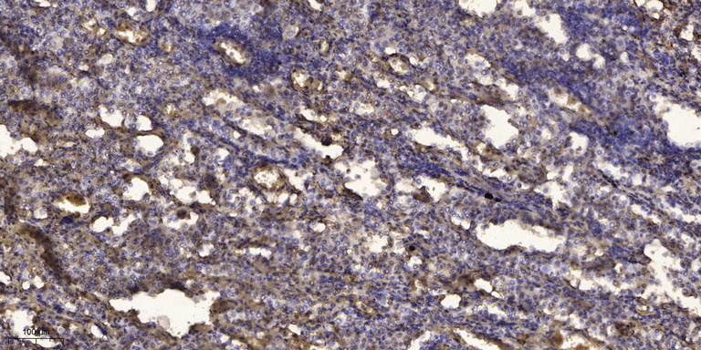
- Immunohistochemical analysis of paraffin-embedded human Squamous cell carcinoma of lung. 1, Antibody was diluted at 1:200(4° overnight). 2, Tris-EDTA,pH9.0 was used for antigen retrieval. 3,Secondary antibody was diluted at 1:200(room temperature, 45min).


