CTNND1 (Phospho Tyr257) Rabbit pAb
- Catalog No.:YP1815
- Applications:IHC;WB
- Reactivity:Human;Mouse
- Target:
- Catenin δ-1
- Fields:
- >>Rap1 signaling pathway;>>Adherens junction;>>Leukocyte transendothelial migration
- Gene Name:
- CTNND1 KIAA0384
- Protein Name:
- Catenin delta-1 (Cadherin-associated Src substrate) (CAS) (p120 catenin) (p120(ctn)) (p120(cas))
- Human Gene Id:
- 1500
- Human Swiss Prot No:
- O60716
- Mouse Gene Id:
- 12388
- Mouse Swiss Prot No:
- P30999
- Immunogen:
- Synthesized peptide derived from human CTNND1 (Phospho Tyr257)
- Specificity:
- This antibody detects endogenous levels of CTNND1 (Phospho Tyr257) Rabbit pAb at Human, Mouse
- Formulation:
- Liquid in PBS containing 50% glycerol, and 0.02% sodium azide.
- Source:
- Rabbit,polyclonal
- Dilution:
- WB 1:500-2000 IHC 1:50-200
- Purification:
- The antibody was affinity-purified from rabbit serum by affinity-chromatography using specific immunogen.
- Concentration:
- 1 mg/ml
- Storage Stability:
- -15°C to -25°C/1 year(Do not lower than -25°C)
- Other Name:
- Catenin delta-1 (Cadherin-associated Src substrate) (CAS) (p120 catenin) (p120(ctn)) (p120(cas))
- Observed Band(KD):
- 108kD
- Background:
- catenin delta 1(CTNND1) Homo sapiens This gene encodes a member of the Armadillo protein family, which function in adhesion between cells and signal transduction. Multiple translation initiation codons and alternative splicing result in many different isoforms being translated. Not all of the full-length natures of the described transcript variants have been determined. Read-through transcription also exists between this gene and the neighboring upstream thioredoxin-related transmembrane protein 2 (TMX2) gene. [provided by RefSeq, Dec 2010],
- Function:
- alternative products:Experimental confirmation may be lacking for some isoforms,disease:May contribute to cell malignancy. Complete loss of expression was observed in approximately 10% of invasive ductal breast carcinomas investigated.,domain:A possible nuclear localization signal exists in all isoforms where Asp-626--631-Arg are deleted.,function:Binds to and inhibits the transcriptional repressor ZBTB33, which may lead to activation of target genes of the Wnt signaling pathway (By similarity). May associate with and regulate the cell adhesion properties of both C- and E-cadherins. Implicated both in cell transformation by SRC and in ligand-induced receptor signaling through the EGF, PDGF, CSF-1 and ERBB2 receptors. Promotes GLIS2 C-terminal cleavage.,induction:Induced in vascular endothelium by wounding. This effect is potentiated by prior laminar shear stress, which enhances wound clo
- Subcellular Location:
- Cell junction, adherens junction . Cytoplasm . Nucleus . Cell membrane . Interaction with GLIS2 promotes nuclear translocation (By similarity). Detected at cell-cell contacts (PubMed:15240885, PubMed:17047063). NANOS1 induces its translocation from sites of cell-cell contact to the cytoplasm (PubMed:17047063). CDH1 enhances cell membrane localization (PubMed:15240885). Isoforms 4A and 1AB are excluded from the nucleus (PubMed:11896187). .; [Isoform 1A]: Nucleus .; [Isoform 2A]: Nucleus .; [Isoform 3A]: Nucleus .
- Expression:
- Expressed in vascular endothelium. Melanocytes and melanoma cells primarily express the long isoform 1A, whereas keratinocytes express shorter isoforms, especially 3A. The shortest isoform 4A, is detected in normal keratinocytes and melanocytes, and generally lost from cells derived from squamous cell carcinomas or melanomas. The C-terminal alternatively spliced exon B is present in the p120ctn transcripts in the colon, intestine and prostate, but lost in several tumor tissues derived from these organs.
- June 19-2018
- WESTERN IMMUNOBLOTTING PROTOCOL
- June 19-2018
- IMMUNOHISTOCHEMISTRY-PARAFFIN PROTOCOL
- June 19-2018
- IMMUNOFLUORESCENCE PROTOCOL
- September 08-2020
- FLOW-CYTOMEYRT-PROTOCOL
- May 20-2022
- Cell-Based ELISA│解您多样本WB检测之困扰
- July 13-2018
- CELL-BASED-ELISA-PROTOCOL-FOR-ACETYL-PROTEIN
- July 13-2018
- CELL-BASED-ELISA-PROTOCOL-FOR-PHOSPHO-PROTEIN
- July 13-2018
- Antibody-FAQs
- Products Images
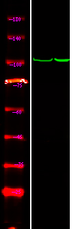
- Western Blot analysis of 1 HepG2 cell, 2 LPS 100ng/mL 30min treated ,using primary antibody at 1:1000 dilution. Secondary antibody(catalog#:RS23920) was diluted at 1:10000


