14-3-3 β Polyclonal Antibody
- Catalog No.:YT0002
- Applications:WB;IHC;IF;ELISA
- Reactivity:Human;Mouse;Rat
- Target:
- 14-3-3 β
- Fields:
- >>Cell cycle;>>Oocyte meiosis;>>PI3K-Akt signaling pathway;>>Hippo signaling pathway;>>Hepatitis C;>>Hepatitis B;>>Viral carcinogenesis
- Gene Name:
- YWHAB
- Protein Name:
- 14-3-3 protein beta/alpha
- Human Gene Id:
- 7529
- Human Swiss Prot No:
- P31946
- Mouse Gene Id:
- 54401
- Mouse Swiss Prot No:
- Q9CQV8
- Rat Gene Id:
- 56011
- Rat Swiss Prot No:
- P35213
- Immunogen:
- The antiserum was produced against synthesized peptide derived from human 14-3-3 beta. AA range:41-90
- Specificity:
- 14-3-3 β Polyclonal Antibody detects endogenous levels of 14-3-3 β protein.
- Formulation:
- Liquid in PBS containing 50% glycerol, 0.5% BSA and 0.02% sodium azide.
- Source:
- Polyclonal, Rabbit,IgG
- Dilution:
- WB 1:500 - 1:2000. IHC 1:100 - 1:300. IF 1:200 - 1:1000. ELISA: 1:10000. Not yet tested in other applications.
- Purification:
- The antibody was affinity-purified from rabbit antiserum by affinity-chromatography using epitope-specific immunogen.
- Concentration:
- 1 mg/ml
- Storage Stability:
- -15°C to -25°C/1 year(Do not lower than -25°C)
- Other Name:
- YWHAB;14-3-3 protein beta/alpha;Protein 1054;Protein kinase C inhibitor protein 1;KCIP-1
- Observed Band(KD):
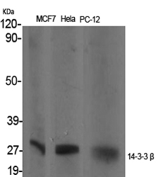
- 28kD
- Background:
- This gene encodes a protein belonging to the 14-3-3 family of proteins, members of which mediate signal transduction by binding to phosphoserine-containing proteins. This highly conserved protein family is found in both plants and mammals. The encoded protein has been shown to interact with RAF1 and CDC25 phosphatases, suggesting that it may play a role in linking mitogenic signaling and the cell cycle machinery. Two transcript variants, which encode the same protein, have been identified for this gene. [provided by RefSeq, Jul 2008],
- Function:
- function:Adapter protein implicated in the regulation of a large spectrum of both general and specialized signaling pathway. Binds to a large number of partners, usually by recognition of a phosphoserine or phosphothreonine motif. Binding generally results in the modulation of the activity of the binding partner. Negative regulator of osteogenesis.,PTM:Isoform Short contains a N-acetylmethionine at position 1.,PTM:The alpha, brain-specific form differs from the beta form in being phosphorylated.,similarity:Belongs to the 14-3-3 family.,subcellular location:Identified by mass spectrometry in melanosome fractions from stage I to stage IV.,subunit:Homodimer. Interacts with SSH1 and TORC2/CRTC2. Interacts with ABL1; the interaction results in cytoplasmic location of ABL1 and inhition of cABL-mediated apoptosis. Interacts with ROR2 (dimer); the interaction results in phosphorylation of YWHAB
- Subcellular Location:
- Cytoplasm . Melanosome . Identified by mass spectrometry in melanosome fractions from stage I to stage IV.; Vacuole membrane . (Microbial infection) Upon infection with Chlamydia trachomatis, this protein is associated with the pathogen-containing vacuole membrane where it colocalizes with IncG. .
- Expression:
- Brain,Colon carcinoma,Kerat
- June 19-2018
- WESTERN IMMUNOBLOTTING PROTOCOL
- June 19-2018
- IMMUNOHISTOCHEMISTRY-PARAFFIN PROTOCOL
- June 19-2018
- IMMUNOFLUORESCENCE PROTOCOL
- September 08-2020
- FLOW-CYTOMEYRT-PROTOCOL
- May 20-2022
- Cell-Based ELISA│解您多样本WB检测之困扰
- July 13-2018
- CELL-BASED-ELISA-PROTOCOL-FOR-ACETYL-PROTEIN
- July 13-2018
- CELL-BASED-ELISA-PROTOCOL-FOR-PHOSPHO-PROTEIN
- July 13-2018
- Antibody-FAQs
- Products Images

- Western Blot analysis of various cells using 14-3-3 β Polyclonal Antibody
.jpg)
- Western Blot analysis of Jurkat cells using 14-3-3 β Polyclonal Antibody

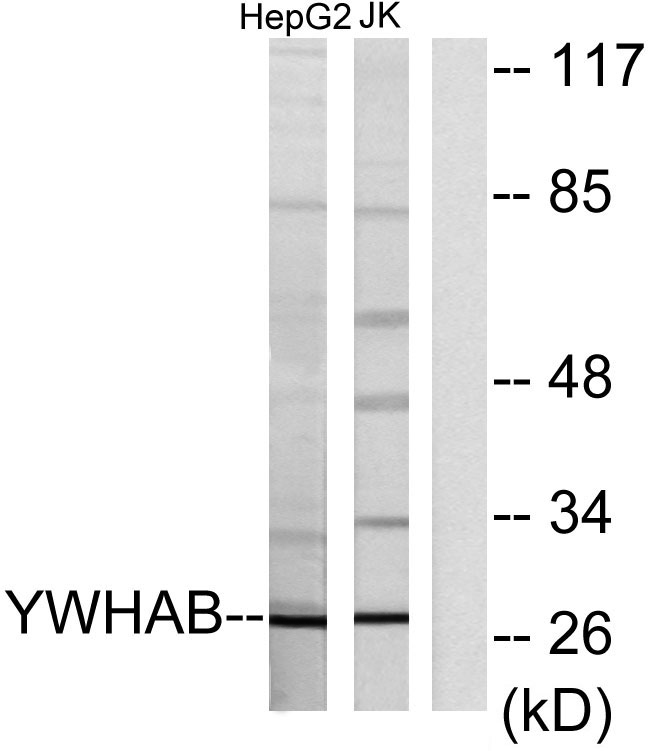
- Western blot analysis of lysates from HepG2 and Jurkat cells, using 14-3-3 beta Antibody. The lane on the right is blocked with the synthesized peptide.

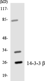
- Western blot analysis of the lysates from HepG2 cells using 14-3-3 β antibody.

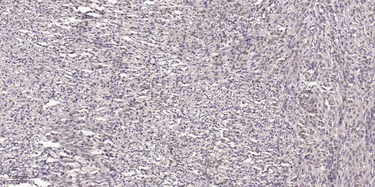
- Immunohistochemical analysis of paraffin-embedded human small intestinal carcinoma tissue. 1,primary Antibody was diluted at 1:200(4° overnight). 2, Sodium citrate pH 6.0 was used for antigen retrieval(>98°C,20min). 3,Secondary antibody was diluted at 1:200


