ApoE Polyclonal Antibody
- Catalog No.:YT0273
- Applications:WB;IHC;IF;ELISA
- Reactivity:Human;Mouse;Rat
- Target:
- ApoE
- Fields:
- >>Cholesterol metabolism;>>Alzheimer disease
- Gene Name:
- APOE
- Protein Name:
- Apolipoprotein E
- Human Gene Id:
- 348
- Human Swiss Prot No:
- P02649
- Mouse Gene Id:
- 11816
- Mouse Swiss Prot No:
- P08226
- Rat Gene Id:
- 25728
- Rat Swiss Prot No:
- P02650
- Immunogen:
- The antiserum was produced against synthesized peptide derived from human ApoE. AA range:37-86
- Specificity:
- ApoE Polyclonal Antibody detects endogenous levels of ApoE protein.
- Formulation:
- Liquid in PBS containing 50% glycerol, 0.5% BSA and 0.02% sodium azide.
- Source:
- Polyclonal, Rabbit,IgG
- Dilution:
- WB 1:500 - 1:2000. IHC 1:100 - 1:300. ELISA: 1:10000.. IF 1:50-200
- Purification:
- The antibody was affinity-purified from rabbit antiserum by affinity-chromatography using epitope-specific immunogen.
- Concentration:
- 1 mg/ml
- Storage Stability:
- -15°C to -25°C/1 year(Do not lower than -25°C)
- Other Name:
- APOE;Apolipoprotein E;Apo-E
- Observed Band(KD):
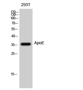
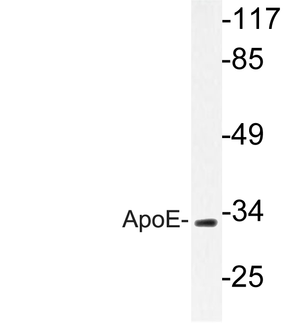
- 36kD
- Background:
- The protein encoded by this gene is a major apoprotein of the chylomicron. It binds to a specific liver and peripheral cell receptor, and is essential for the normal catabolism of triglyceride-rich lipoprotein constituents. This gene maps to chromosome 19 in a cluster with the related apolipoprotein C1 and C2 genes. Mutations in this gene result in familial dysbetalipoproteinemia, or type III hyperlipoproteinemia (HLP III), in which increased plasma cholesterol and triglycerides are the consequence of impaired clearance of chylomicron and VLDL remnants. [provided by RefSeq, Jun 2016],
- Function:
- disease:Defects in APOE are a cause of hyperlipoproteinemia type III [MIM:107741]; also known as familial dysbetalipoproteinemia. Individuals with hyperlipoproteinemia type III, are clinically characterized by xanthomas, yellowish lipid deposits in the palmar crease, or less specific on tendons and on elbows. The disorder rarely manifests before the third decade in men. In women, it is usually expressed only after the menopause. The vast majority of the patients are homozygous for APOE*2 alleles. More severe cases of hyperlipoproteinemia type III have also been observed in individuals heterozygous for rare APOE variants. The influence of APOE on lipid levels is often suggested to have major implications for the risk of coronary artery disease (CAD). Individuals carrying the common APOE*4 variant are at higher risk of CAD.,disease:Defects in APOE are a cause of lipoprotein glomerulopathy
- Subcellular Location:
- Secreted . Secreted, extracellular space . Secreted, extracellular space, extracellular matrix . In the plasma, APOE is associated with chylomicrons, chylomicrons remnants, VLDL, LDL and HDL lipoproteins (PubMed:1911868, PubMed:8340399). Lipid poor oligomeric APOE is associated with the extracellular matrix in a calcium- and heparan-sulfate proteoglycans-dependent manner (PubMed:9488694). Lipidation induces the release from the extracellular matrix (PubMed:9488694). .
- Expression:
- Produced by several tissues and cell types and mainly found associated with lipid particles in the plasma, the interstitial fluid and lymph (PubMed:25173806). Mainly synthesized by liver hepatocytes (PubMed:25173806). Significant quantities are also produced in brain, mainly by astrocytes and glial cells in the cerebral cortex, but also by neurons in frontal cortex and hippocampus (PubMed:3115992, PubMed:10027417). It is also expressed by cells of the peripheral nervous system (PubMed:10027417, PubMed:25173806). Also expressed by adrenal gland, testis, ovary, skin, kidney, spleen and adipose tissue and macrophages in various tissues (PubMed:25173806).
Quantitative dot blot analysis (QDB), a versatile high throughput immunoblot method. Oncotarget Oncotarget. 2017 Aug 29; 8(35): 58553–58562 WB Human liver HEK293 cell
Zhang, Jiandi. "Apparatus and method for high trhoughput immunobloting." U.S. Patent Application No. 15/433,586.
- June 19-2018
- WESTERN IMMUNOBLOTTING PROTOCOL
- June 19-2018
- IMMUNOHISTOCHEMISTRY-PARAFFIN PROTOCOL
- June 19-2018
- IMMUNOFLUORESCENCE PROTOCOL
- September 08-2020
- FLOW-CYTOMEYRT-PROTOCOL
- May 20-2022
- Cell-Based ELISA│解您多样本WB检测之困扰
- July 13-2018
- CELL-BASED-ELISA-PROTOCOL-FOR-ACETYL-PROTEIN
- July 13-2018
- CELL-BASED-ELISA-PROTOCOL-FOR-PHOSPHO-PROTEIN
- July 13-2018
- Antibody-FAQs
- Products Images

- Western Blot analysis of 293T cells using ApoE Polyclonal Antibody diluted at 1:500

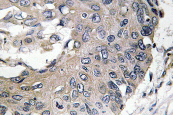
- Immunohistochemistry analysis of ApoE antibody in paraffin-embedded human lung carcinoma tissue.

- Western blot analysis of lysate from RAW264.7cells, using ApoE antibody.


