BART1 Polyclonal Antibody
- Catalog No.:YT0453
- Applications:WB;ELISA
- Reactivity:Human;Mouse;Rat
- Target:
- BART1
- Gene Name:
- ARL2BP
- Protein Name:
- ADP-ribosylation factor-like protein 2-binding protein
- Human Gene Id:
- 23568
- Human Swiss Prot No:
- Q9Y2Y0
- Mouse Gene Id:
- 107566
- Mouse Swiss Prot No:
- Q9D385
- Rat Gene Id:
- 498910
- Rat Swiss Prot No:
- Q4V8C5
- Immunogen:
- The antiserum was produced against synthesized peptide derived from human ARL2BP. AA range:101-150
- Specificity:
- BART1 Polyclonal Antibody detects endogenous levels of BART1 protein.
- Formulation:
- Liquid in PBS containing 50% glycerol, 0.5% BSA and 0.02% sodium azide.
- Source:
- Polyclonal, Rabbit,IgG
- Dilution:
- WB 1:500 - 1:2000. ELISA: 1:20000. Not yet tested in other applications.
- Purification:
- The antibody was affinity-purified from rabbit antiserum by affinity-chromatography using epitope-specific immunogen.
- Concentration:
- 1 mg/ml
- Storage Stability:
- -15°C to -25°C/1 year(Do not lower than -25°C)
- Other Name:
- ARL2BP;BART;BART1;ADP-ribosylation factor-like protein 2-binding protein;ARF-like 2-binding protein;Binder of ARF2 protein 1
- Observed Band(KD):
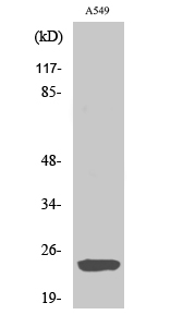
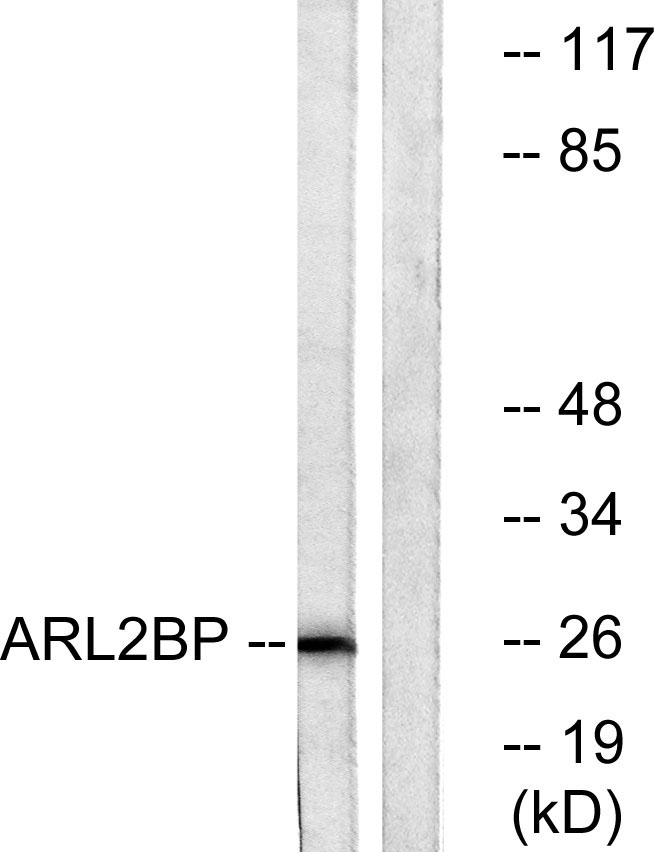
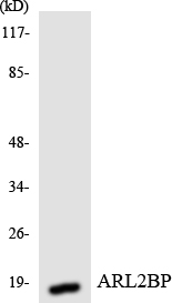
- 25kD
- Background:
- ADP-ribosylation factor (ARF)-like proteins (ARLs) comprise a functionally distinct group of the ARF family of RAS-related GTPases. The protein encoded by this gene binds to ARL2.GTP with high affinity but does not interact with ARL2.GDP, activated ARF, or RHO proteins. The lack of detectable membrane association of this protein or ARL2 upon activation of ARL2 is suggestive of actions distinct from those of the ARFs. This protein is considered to be the first ARL2-specific effector identified, due to its interaction with ARL2.GTP but lack of ARL2 GTPase-activating protein activity. [provided by RefSeq, Jul 2008],
- Function:
- function:May play a role as an effector of the ADP-ribosylation factor-like protein 2, ARL2.,PTM:Phosphorylated upon DNA damage, probably by ATM or ATR.,similarity:Belongs to the ARL2BP family.,subunit:Interacts with GTP bound ARL2 and ARL3; the complex ARL2-ARL2BP as well as ARL2BP alone, binds to ANT1.,tissue specificity:Ubiquitous.,
- Subcellular Location:
- Cytoplasm. Mitochondrion intermembrane space. Cytoplasm, cytoskeleton, microtubule organizing center, centrosome. Nucleus. Cytoplasm, cytoskeleton, spindle. Cytoplasm, cytoskeleton, cilium basal body. The complex formed with ARL2BP, ARL2 and SLC25A4 is expressed in mitochondria (By similarity). Detected in the midbody matrix. Not detected in the Golgi, nucleus and on the mitotic spindle. Centrosome-associated throughout the cell cycle. Not detected to interphase microtubules. In retina photoreceptor cells, localized in the distal connecting cilia, basal body, ciliary-associated centriole, and ciliary rootlet. Interaction with ARL2 may be required for cilia basal body localization. .
- Expression:
- Expressed in retina pigment epithelial cells (at protein level). Widely expressed.
- June 19-2018
- WESTERN IMMUNOBLOTTING PROTOCOL
- June 19-2018
- IMMUNOHISTOCHEMISTRY-PARAFFIN PROTOCOL
- June 19-2018
- IMMUNOFLUORESCENCE PROTOCOL
- September 08-2020
- FLOW-CYTOMEYRT-PROTOCOL
- May 20-2022
- Cell-Based ELISA│解您多样本WB检测之困扰
- July 13-2018
- CELL-BASED-ELISA-PROTOCOL-FOR-ACETYL-PROTEIN
- July 13-2018
- CELL-BASED-ELISA-PROTOCOL-FOR-PHOSPHO-PROTEIN
- July 13-2018
- Antibody-FAQs
- Products Images

- Western Blot analysis of various cells using BART1 Polyclonal Antibody

- Western blot analysis of lysates from A549 cells, using ARL2BP Antibody. The lane on the right is blocked with the synthesized peptide.

- Western blot analysis of the lysates from HUVECcells using ARL2BP antibody.


