C/EBP β Polyclonal Antibody
- Catalog No.:YT0553
- Applications:IF;WB;IHC;ELISA
- Reactivity:Human;Mouse;Rat;Pig
- Target:
- C/EBP β
- Fields:
- >>IL-17 signaling pathway;>>TNF signaling pathway;>>Tuberculosis;>>Transcriptional misregulation in cancer
- Gene Name:
- CEBPB
- Protein Name:
- CCAAT/enhancer-binding protein beta
- Human Gene Id:
- 1051
- Human Swiss Prot No:
- P17676
- Mouse Gene Id:
- 12608
- Mouse Swiss Prot No:
- P28033
- Rat Gene Id:
- 24253
- Rat Swiss Prot No:
- P21272
- Immunogen:
- The antiserum was produced against synthesized peptide derived from human C/EBP-beta. AA range:201-250
- Specificity:
- C/EBP β Polyclonal Antibody detects endogenous levels of C/EBP β protein.
- Formulation:
- Liquid in PBS containing 50% glycerol, 0.5% BSA and 0.02% sodium azide.
- Source:
- Polyclonal, Rabbit,IgG
- Dilution:
- IF 1:50-200 WB 1:500 - 1:2000. IHC 1:100 - 1:300. ELISA: 1:10000. Not yet tested in other applications.
- Purification:
- The antibody was affinity-purified from rabbit antiserum by affinity-chromatography using epitope-specific immunogen.
- Concentration:
- 1 mg/ml
- Storage Stability:
- -15°C to -25°C/1 year(Do not lower than -25°C)
- Other Name:
- CEBPB;LAP;TCF5;PP9092;CCAAT/enhancer-binding protein beta;C/EBP beta;Liver activator protein;Nuclear factor NF-IL6;Transcription factor 5;TCF-5
- Observed Band(KD):
- 36kD
- Background:
- This intronless gene encodes a transcription factor that contains a basic leucine zipper (bZIP) domain. The encoded protein functions as a homodimer but can also form heterodimers with CCAAT/enhancer-binding proteins alpha, delta, and gamma. Activity of this protein is important in the regulation of genes involved in immune and inflammatory responses, among other processes. The use of alternative in-frame AUG start codons results in multiple protein isoforms, each with distinct biological functions. [provided by RefSeq, Oct 2013],
- Function:
- function:Important transcriptional activator in the regulation of genes involved in immune and inflammatory responses. Specifically binds to an IL-1 response element in the IL-6 gene. NF-IL6 also binds to regulatory regions of several acute-phase and cytokines genes. It probably plays a role in the regulation of acute-phase reaction, inflammation and hemopoiesis. The consensus recognition site is 5'-T[TG]NNGNAA[TG]-3'.,PTM:Sumoylated by polymeric chains of SUMO2 or SUMO3.,similarity:Belongs to the bZIP family.,similarity:Belongs to the bZIP family. C/EBP subfamily.,similarity:Contains 1 bZIP domain.,subunit:Binds DNA as a dimer and can form stable heterodimers with C/EBP alpha, delta and gamma. Interacts with TRIM28 and PTGES2.,tissue specificity:Expressed at low levels in the lung, kidney and spleen.,
- Subcellular Location:
- Nucleus . Cytoplasm . Translocates to the nucleus when phosphorylated at Ser-288. In T-cells when sumoylated drawn to pericentric heterochromatin thereby allowing proliferation (By similarity). .
- Expression:
- Expressed at low levels in the lung, kidney and spleen.
The C/EBPβ-Dependent Induction of TFDP2 Facilitates Porcine Reproductive and Respiratory Syndrome Virus Proliferation. VIROLOGICA SINICA Virol Sin. 2021 Dec;36(6):1341-1351 WB Pig 1:1000 3D4/21 cells
- June 19-2018
- WESTERN IMMUNOBLOTTING PROTOCOL
- June 19-2018
- IMMUNOHISTOCHEMISTRY-PARAFFIN PROTOCOL
- June 19-2018
- IMMUNOFLUORESCENCE PROTOCOL
- September 08-2020
- FLOW-CYTOMEYRT-PROTOCOL
- May 20-2022
- Cell-Based ELISA│解您多样本WB检测之困扰
- July 13-2018
- CELL-BASED-ELISA-PROTOCOL-FOR-ACETYL-PROTEIN
- July 13-2018
- CELL-BASED-ELISA-PROTOCOL-FOR-PHOSPHO-PROTEIN
- July 13-2018
- Antibody-FAQs
- Products Images
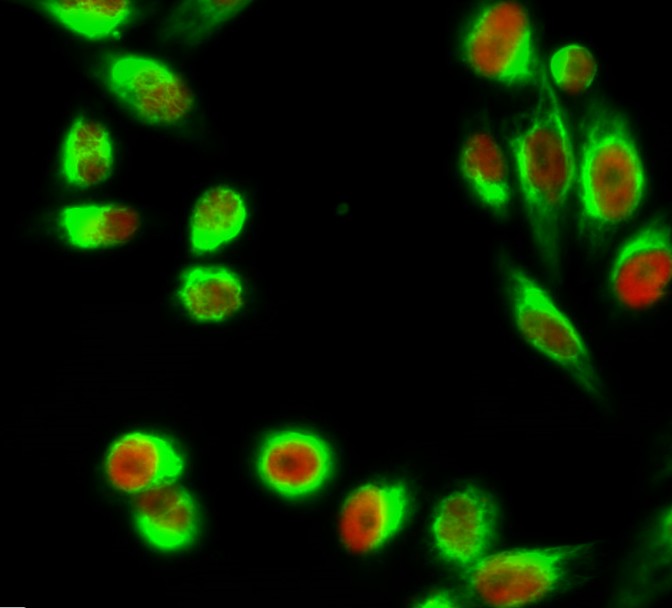
- Immunofluorescence analysis of Hela cell. 1,C/EBP β Polyclonal Antibody(red) was diluted at 1:200(4° overnight). Kif 7 Monoclonal Antibody(3F8)(green) was diluted at 1:200(4° overnight). 2, Goat Anti Rabbit Alexa Fluor 594 Catalog:RS3611 was diluted at 1:1000(room temperature, 50min). Goat Anti Mouse Alexa Fluor 488 Catalog:RS3208 was diluted at 1:1000(room temperature, 50min).
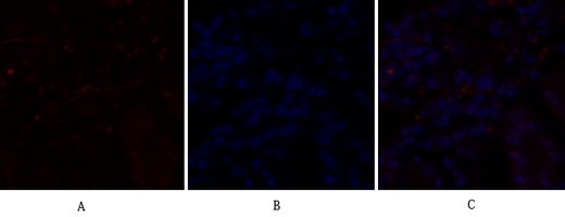
- Immunofluorescence analysis of human-stomach tissue. 1,C/EBP β Polyclonal Antibody(red) was diluted at 1:200(4°C,overnight). 2, Cy3 labled Secondary antibody was diluted at 1:300(room temperature, 50min).3, Picture B: DAPI(blue) 10min. Picture A:Target. Picture B: DAPI. Picture C: merge of A+B
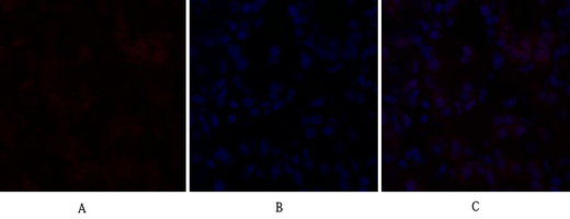
- Immunofluorescence analysis of human-stomach tissue. 1,C/EBP β Polyclonal Antibody(red) was diluted at 1:200(4°C,overnight). 2, Cy3 labled Secondary antibody was diluted at 1:300(room temperature, 50min).3, Picture B: DAPI(blue) 10min. Picture A:Target. Picture B: DAPI. Picture C: merge of A+B
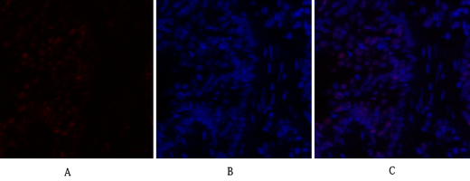
- Immunofluorescence analysis of rat-lung tissue. 1,C/EBP β Polyclonal Antibody(red) was diluted at 1:200(4°C,overnight). 2, Cy3 labled Secondary antibody was diluted at 1:300(room temperature, 50min).3, Picture B: DAPI(blue) 10min. Picture A:Target. Picture B: DAPI. Picture C: merge of A+B
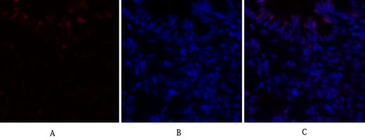
- Immunofluorescence analysis of rat-lung tissue. 1,C/EBP β Polyclonal Antibody(red) was diluted at 1:200(4°C,overnight). 2, Cy3 labled Secondary antibody was diluted at 1:300(room temperature, 50min).3, Picture B: DAPI(blue) 10min. Picture A:Target. Picture B: DAPI. Picture C: merge of A+B
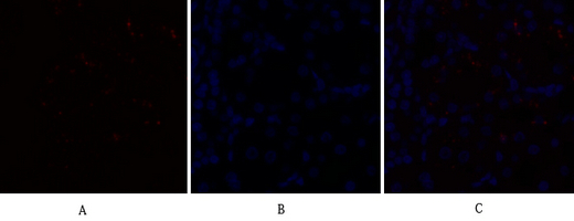
- Immunofluorescence analysis of rat-kidney tissue. 1,C/EBP β Polyclonal Antibody(red) was diluted at 1:200(4°C,overnight). 2, Cy3 labled Secondary antibody was diluted at 1:300(room temperature, 50min).3, Picture B: DAPI(blue) 10min. Picture A:Target. Picture B: DAPI. Picture C: merge of A+B
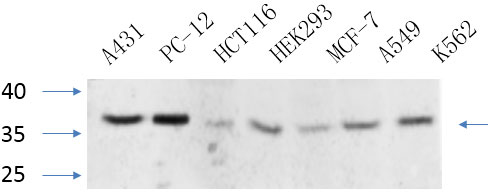
- Western Blot analysis of various cells using primary antibody diluted at 1:1000(4°C overnight). Secondary antibody:Goat Anti-rabbit IgG IRDye 800( diluted at 1:5000, 25°C, 1 hour). Cell lysate was extracted by Minute™ Plasma Membrane Protein Isolation and Cell Fractionation Kit(SM-005, Inventbiotech,MN,USA).
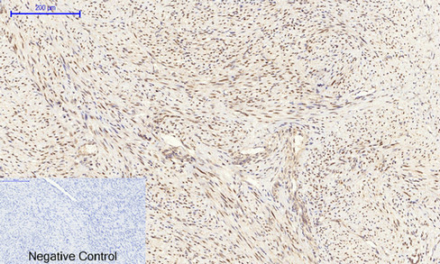
- Immunohistochemical analysis of paraffin-embedded Human-uterus-cancer tissue. 1,C/EBP β Polyclonal Antibody was diluted at 1:200(4°C,overnight). 2, Sodium citrate pH 6.0 was used for antibody retrieval(>98°C,20min). 3,Secondary antibody was diluted at 1:200(room tempeRature, 30min). Negative control was used by secondary antibody only.
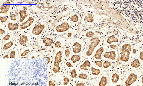
- Immunohistochemical analysis of paraffin-embedded Human-stomach tissue. 1,C/EBP β Polyclonal Antibody was diluted at 1:200(4°C,overnight). 2, Sodium citrate pH 6.0 was used for antibody retrieval(>98°C,20min). 3,Secondary antibody was diluted at 1:200(room tempeRature, 30min). Negative control was used by secondary antibody only.
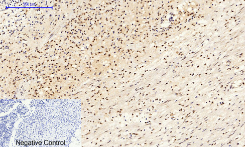
- Immunohistochemical analysis of paraffin-embedded Human-Appendix tissue. 1,C/EBP β Polyclonal Antibody was diluted at 1:200(4°C,overnight). 2, Sodium citrate pH 6.0 was used for antibody retrieval(>98°C,20min). 3,Secondary antibody was diluted at 1:200(room tempeRature, 30min). Negative control was used by secondary antibody only.
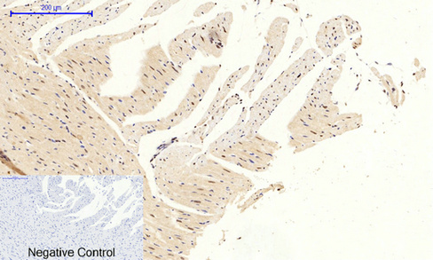
- Immunohistochemical analysis of paraffin-embedded Rat-heart tissue. 1,C/EBP β Polyclonal Antibody was diluted at 1:200(4°C,overnight). 2, Sodium citrate pH 6.0 was used for antibody retrieval(>98°C,20min). 3,Secondary antibody was diluted at 1:200(room tempeRature, 30min). Negative control was used by secondary antibody only.
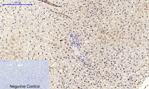
- Immunohistochemical analysis of paraffin-embedded Rat-liver tissue. 1,C/EBP β Polyclonal Antibody was diluted at 1:200(4°C,overnight). 2, Sodium citrate pH 6.0 was used for antibody retrieval(>98°C,20min). 3,Secondary antibody was diluted at 1:200(room tempeRature, 30min). Negative control was used by secondary antibody only.
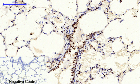
- Immunohistochemical analysis of paraffin-embedded Rat-lung tissue. 1,C/EBP β Polyclonal Antibody was diluted at 1:200(4°C,overnight). 2, Sodium citrate pH 6.0 was used for antibody retrieval(>98°C,20min). 3,Secondary antibody was diluted at 1:200(room tempeRature, 30min). Negative control was used by secondary antibody only.
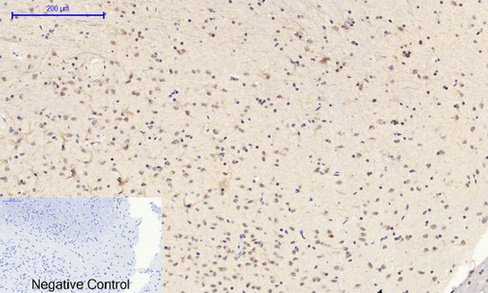
- Immunohistochemical analysis of paraffin-embedded Rat-brain tissue. 1,C/EBP β Polyclonal Antibody was diluted at 1:200(4°C,overnight). 2, Sodium citrate pH 6.0 was used for antibody retrieval(>98°C,20min). 3,Secondary antibody was diluted at 1:200(room tempeRature, 30min). Negative control was used by secondary antibody only.
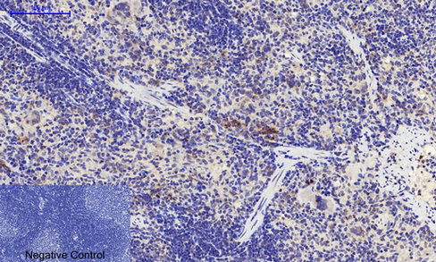
- Immunohistochemical analysis of paraffin-embedded Rat-spleen tissue. 1,C/EBP β Polyclonal Antibody was diluted at 1:200(4°C,overnight). 2, Sodium citrate pH 6.0 was used for antibody retrieval(>98°C,20min). 3,Secondary antibody was diluted at 1:200(room tempeRature, 30min). Negative control was used by secondary antibody only.
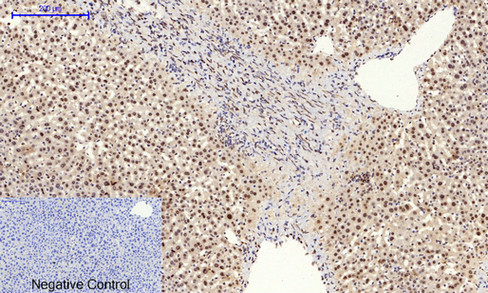
- Immunohistochemical analysis of paraffin-embedded Mouse-liver tissue. 1,C/EBP β Polyclonal Antibody was diluted at 1:200(4°C,overnight). 2, Sodium citrate pH 6.0 was used for antibody retrieval(>98°C,20min). 3,Secondary antibody was diluted at 1:200(room tempeRature, 30min). Negative control was used by secondary antibody only.
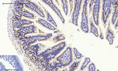
- Immunohistochemical analysis of paraffin-embedded Mouse-colon tissue. 1,C/EBP β Polyclonal Antibody was diluted at 1:200(4°C,overnight). 2, Sodium citrate pH 6.0 was used for antibody retrieval(>98°C,20min). 3,Secondary antibody was diluted at 1:200(room tempeRature, 30min). Negative control was used by secondary antibody only.
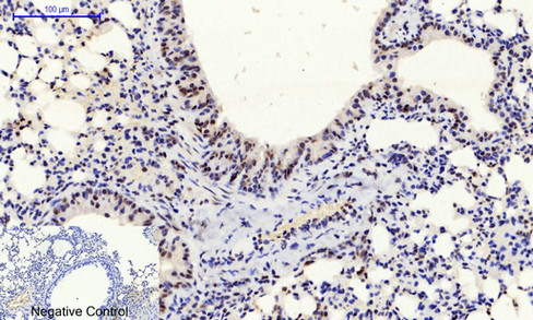
- Immunohistochemical analysis of paraffin-embedded Mouse-lung tissue. 1,C/EBP β Polyclonal Antibody was diluted at 1:200(4°C,overnight). 2, Sodium citrate pH 6.0 was used for antibody retrieval(>98°C,20min). 3,Secondary antibody was diluted at 1:200(room tempeRature, 30min). Negative control was used by secondary antibody only.
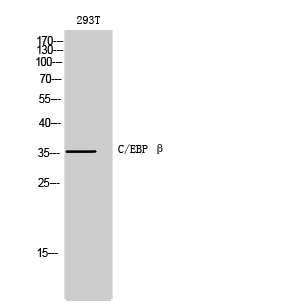
- Western Blot analysis of 293T cells using C/EBP β Polyclonal Antibody diluted at 1:500
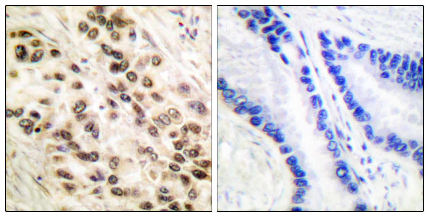
- Immunohistochemistry analysis of paraffin-embedded human lung carcinoma tissue, using C/EBP-beta Antibody. The picture on the right is blocked with the synthesized peptide.
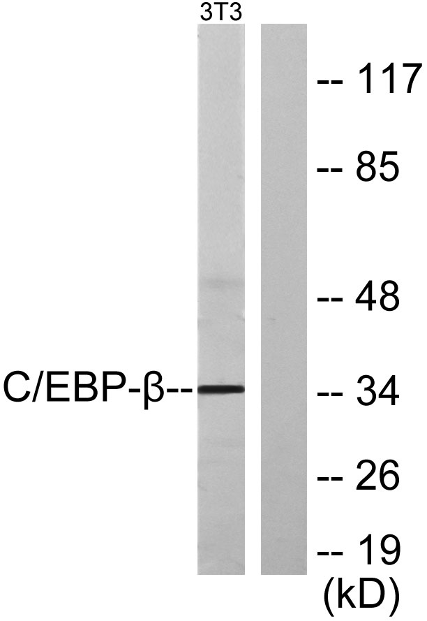
- Western blot analysis of lysates from NIH/3T3 cells, using C/EBP-beta Antibody. The lane on the right is blocked with the synthesized peptide.


