Calnexin Polyclonal Antibody
- Catalog No.:YT0613
- Applications:WB;IHC;IF;ELISA
- Reactivity:Human;Mouse;Rat
- Target:
- Calnexin
- Fields:
- >>Protein processing in endoplasmic reticulum;>>Phagosome;>>Antigen processing and presentation;>>Thyroid hormone synthesis;>>Human T-cell leukemia virus 1 infection
- Gene Name:
- CANX
- Protein Name:
- Calnexin
- Human Gene Id:
- 821
- Human Swiss Prot No:
- P27824
- Mouse Gene Id:
- 12330
- Mouse Swiss Prot No:
- P35564
- Rat Gene Id:
- 29144
- Rat Swiss Prot No:
- P35565
- Immunogen:
- The antiserum was produced against synthesized peptide derived from human Calnexin. AA range:543-592
- Specificity:
- Calnexin Polyclonal Antibody detects endogenous levels of Calnexin protein.
- Formulation:
- Liquid in PBS containing 50% glycerol, 0.5% BSA and 0.02% sodium azide.
- Source:
- Polyclonal, Rabbit,IgG
- Dilution:
- WB 1:500 - 1:2000. IHC: 1:100-300 ELISA: 1:20000. IF 1:100-300 Not yet tested in other applications.
- Purification:
- The antibody was affinity-purified from rabbit antiserum by affinity-chromatography using epitope-specific immunogen.
- Concentration:
- 1 mg/ml
- Storage Stability:
- -15°C to -25°C/1 year(Do not lower than -25°C)
- Other Name:
- CANX;Calnexin;IP90;Major histocompatibility complex class I antigen-binding protein p88;p90
- Observed Band(KD):
- 90kD
- Background:
- This gene encodes a member of the calnexin family of molecular chaperones. The encoded protein is a calcium-binding, endoplasmic reticulum (ER)-associated protein that interacts transiently with newly synthesized N-linked glycoproteins, facilitating protein folding and assembly. It may also play a central role in the quality control of protein folding by retaining incorrectly folded protein subunits within the ER for degradation. Alternatively spliced transcript variants encoding the same protein have been described. [provided by RefSeq, Jul 2008],
- Function:
- function:Calcium-binding protein that interacts with newly synthesized glycoproteins in the endoplasmic reticulum. It may act in assisting protein assembly and/or in the retention within the ER of unassembled protein subunits. It seems to play a major role in the quality control apparatus of the ER by the retention of incorrectly folded proteins.,online information:Calnexin entry,similarity:Belongs to the calreticulin family.,subcellular location:Identified by mass spectrometry in melanosome fractions from stage I to stage IV.,
- Subcellular Location:
- Endoplasmic reticulum membrane ; Single-pass type I membrane protein . Endoplasmic reticulum . Melanosome . Identified by mass spectrometry in melanosome fractions from stage I to stage IV (PubMed:12643545, PubMed:17081065). The palmitoylated form preferentially localizes to the perinuclear rough ER (PubMed:22314232). .
- Expression:
- B-cell lymphoma,Epithelium,Fibroblast,Keratinocyte,Kidney,Liver,Lymph,Placenta,Platelet,
Phenotypic profiling of pancreatic ductal adenocarcinoma plasma-derived small extracellular vesicles for cancer diagnosis and cancer stage prediction: a proof-of-concept study WB Human /sEVs
Periodontal ligament fibroblasts regulate osteoblasts by exosome secretion induced by inflammatory stimuli. ARCHIVES OF ORAL BIOLOGY Arch Oral Biol. 2019 Sep;105:27 WB Human 1:1000 hPDLFs
CD44-targeting Drug Delivery System of Exosomes Loading Forsythiaside A Combats Liver Fibrosis via Regulating NLRP3-mediated Pyroptosis Advanced Healthcare Materials Cheng Peng WB Bovine exosomes
Phillygenin inhibited M1 macrophage polarization and reduced hepatic stellate cell activation by inhibiting macrophage exosomal miR-125b-5p BIOMEDICINE & PHARMACOTHERAPY Cheng Ma WB Mouse RAW264.7 cells
New Findings: Hindlimb Unloading Causes Nucleocytoplasmic Ca2+ Overload and DNA Damage in Skeletal Muscle Cells Yunfang Gao WB Rat muscle cell
Effects of Plasma-Derived Exosomal miRNA-19b-3p on Treg/T Helper 17 Cell Imbalance in Behçet's Uveitis INVESTIGATIVE OPHTHALMOLOGY & VISUAL SCIENCE Peizeng Yang WB Human Exosome
Allogeneic Adipose-Derived Mesenchymal Stem Cell Transplantation Alleviates Atherosclerotic Plaque by Inhibiting Ox-LDL Uptake, Inflammatory Reaction and Endothelial Damage in Rabbits. Chuan Qin WB Rabbit rabbit Adipose-derived stem cells (ADSCs)
Preparation of Phillygenin-Hyaluronic acid composite milk-derived exosomes and its anti-hepatic fibrosis effect. Materials Today Bio Yunxia Li WB Bovine 1:1000 Milk-derived exosomes (mEXO)
Proteomic profiling of aqueous humor-derived exosomes in Vogt-Koyanagi-Harada disease and Behcet's uveitis CLINICAL IMMUNOLOGY Yinan Zhang WB Human 1:1000 aqueous humor (AH) exosome
Elevated extracellular matrix protein 1 in circulating extracellular vesicles supports breast cancer progression under obesity conditions Nature Communications Xu Keyang WB Human 1:500 small extracellular vesicles (sEVs)
Effect of plasma exosome lncRNA on isoproterenol hydrochloride-induced cardiotoxicity in rats TOXICOLOGY AND APPLIED PHARMACOLOGY Liyuan Zhao WB Rat 1:1000 exosome
Multi-platform omics sequencing dissects the atlas of plasma-derived exosomes in rats with or without depression-like behavior after traumatic spinal cord injury PROGRESS IN NEURO-PSYCHOPHARMACOLOGY & BIOLOGICAL PSYCHIATRY Zhihua Wang WB Rat exosomes
Intraocular pressure across the lifespan of Tg-MYOCY437H mice EXPERIMENTAL EYE RESEARCH Xiaoyan Zhang IHC Mouse 1:200 eye
Adipose-Derived exosome from Diet-Induced-Obese mouse attenuates LPS-Induced acute lung injury by inhibiting inflammation and Apoptosis: In vivo and in silico insight INTERNATIONAL IMMUNOPHARMACOLOGY Fengyuan Wang WB Mouse exosome
- June 19-2018
- WESTERN IMMUNOBLOTTING PROTOCOL
- June 19-2018
- IMMUNOHISTOCHEMISTRY-PARAFFIN PROTOCOL
- June 19-2018
- IMMUNOFLUORESCENCE PROTOCOL
- September 08-2020
- FLOW-CYTOMEYRT-PROTOCOL
- May 20-2022
- Cell-Based ELISA│解您多样本WB检测之困扰
- July 13-2018
- CELL-BASED-ELISA-PROTOCOL-FOR-ACETYL-PROTEIN
- July 13-2018
- CELL-BASED-ELISA-PROTOCOL-FOR-PHOSPHO-PROTEIN
- July 13-2018
- Antibody-FAQs
- Products Images
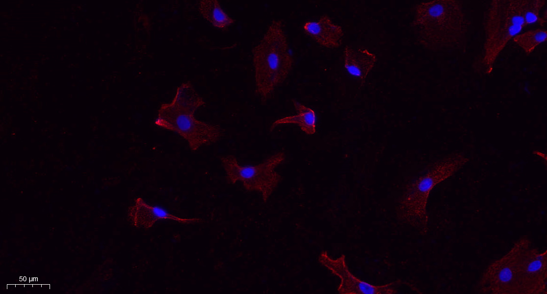
- Immunofluorescence analysis of A549. 1,primary Antibody(red) was diluted at 1:200(4°C overnight). 2, Goat Anti Rabbit IgG (H&L) - Alexa Fluor 594 Secondary antibody was diluted at 1:1000(room temperature, 50min).3, Picture B: DAPI(blue) 10min.
.jpg)
- Zhang, Z., Xu, R., Yang, Y. et al. Micro/nano-textured hierarchical titanium topography promotes exosome biogenesis and secretion to improve osseointegration. J Nanobiotechnol 19, 78 (2021).
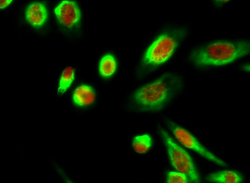
- Immunofluorescence analysis of Hela cell. 1,Calnexin Polyclonal Antibody(green) was diluted at 1:200(4° overnight). (red) was diluted at 1:200(4° overnight). 2, Goat Anti Rabbit Alexa Fluor 488 Catalog:RS3211 was diluted at 1:1000(room temperature, 50min). Goat Anti Mouse Alexa Fluor 594 Catalog:RS3608 was diluted at 1:1000(room temperature, 50min).
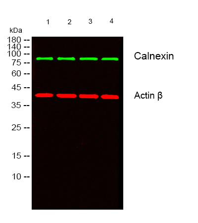
- Western blot analysis of lysates from 1) HCT116, 2) HEK293, 3) MCF-7, 4) A549 cells, (Green) primary antibody was diluted at 1:1000, 4°over night, secondary antibody(cat:RS23920)was diluted at 1:10000, 37° 1hour. (Red) Actin β Monoclonal Antibody(5B7) (cat:YM3028) antibody was diluted at 1:5000 as loading control, 4° over night,secondary antibody(cat:RS23710)was diluted at 1:10000, 37° 1hour.

- Western blot analysis of lysates from HT-29, NIH/3T3, and HepG2 cells, primary antibody was diluted at 1:1000, 4° over night, secondary antibody(cat: RS23920)was diluted at 1:10000, 37° 1hour.
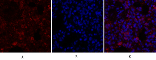
- Immunofluorescence analysis of rat-lung tissue. 1,Calnexin Polyclonal Antibody(red) was diluted at 1:200(4°C,overnight). 2, Cy3 labled Secondary antibody was diluted at 1:300(room temperature, 50min).3, Picture B: DAPI(blue) 10min. Picture A:Target. Picture B: DAPI. Picture C: merge of A+B
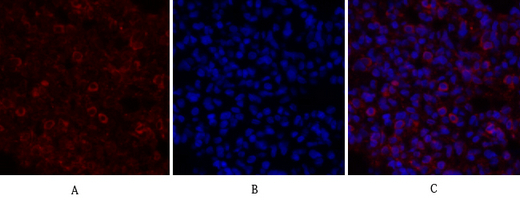
- Immunofluorescence analysis of rat-lung tissue. 1,Calnexin Polyclonal Antibody(red) was diluted at 1:200(4°C,overnight). 2, Cy3 labled Secondary antibody was diluted at 1:300(room temperature, 50min).3, Picture B: DAPI(blue) 10min. Picture A:Target. Picture B: DAPI. Picture C: merge of A+B

- Immunofluorescence analysis of rat-kidney tissue. 1,Calnexin Polyclonal Antibody(red) was diluted at 1:200(4°C,overnight). 2, Cy3 labled Secondary antibody was diluted at 1:300(room temperature, 50min).3, Picture B: DAPI(blue) 10min. Picture A:Target. Picture B: DAPI. Picture C: merge of A+B
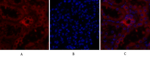
- Immunofluorescence analysis of rat-kidney tissue. 1,Calnexin Polyclonal Antibody(red) was diluted at 1:200(4°C,overnight). 2, Cy3 labled Secondary antibody was diluted at 1:300(room temperature, 50min).3, Picture B: DAPI(blue) 10min. Picture A:Target. Picture B: DAPI. Picture C: merge of A+B
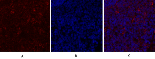
- Immunofluorescence analysis of rat-spleen tissue. 1,Calnexin Polyclonal Antibody(red) was diluted at 1:200(4°C,overnight). 2, Cy3 labled Secondary antibody was diluted at 1:300(room temperature, 50min).3, Picture B: DAPI(blue) 10min. Picture A:Target. Picture B: DAPI. Picture C: merge of A+B
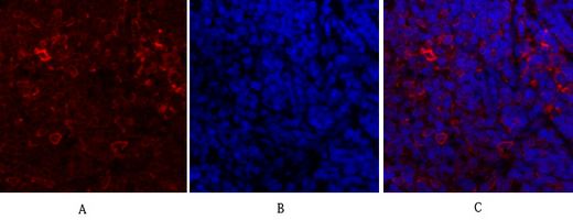
- Immunofluorescence analysis of rat-spleen tissue. 1,Calnexin Polyclonal Antibody(red) was diluted at 1:200(4°C,overnight). 2, Cy3 labled Secondary antibody was diluted at 1:300(room temperature, 50min).3, Picture B: DAPI(blue) 10min. Picture A:Target. Picture B: DAPI. Picture C: merge of A+B
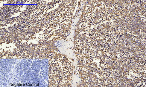
- Immunohistochemical analysis of paraffin-embedded Human-Tonsil tissue. 1,Calnexin Polyclonal Antibody was diluted at 1:200(4°C,overnight). 2, Sodium citrate pH 6.0 was used for antibody retrieval(>98°C,20min). 3,Secondary antibody was diluted at 1:200(room tempeRature, 30min). Negative control was used by secondary antibody only.
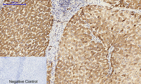
- Immunohistochemical analysis of paraffin-embedded Human-liver tissue. 1,Calnexin Polyclonal Antibody was diluted at 1:200(4°C,overnight). 2, Sodium citrate pH 6.0 was used for antibody retrieval(>98°C,20min). 3,Secondary antibody was diluted at 1:200(room tempeRature, 30min). Negative control was used by secondary antibody only.
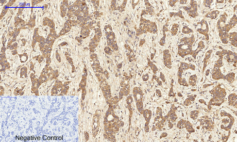
- Immunohistochemical analysis of paraffin-embedded Human-liver-cancer tissue. 1,Calnexin Polyclonal Antibody was diluted at 1:200(4°C,overnight). 2, Sodium citrate pH 6.0 was used for antibody retrieval(>98°C,20min). 3,Secondary antibody was diluted at 1:200(room tempeRature, 30min). Negative control was used by secondary antibody only.
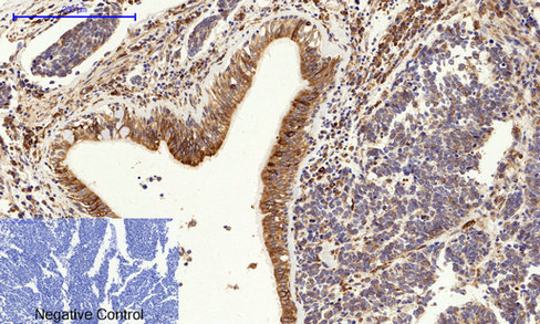
- Immunohistochemical analysis of paraffin-embedded Human-lung-cancer tissue. 1,Calnexin Polyclonal Antibody was diluted at 1:200(4°C,overnight). 2, Sodium citrate pH 6.0 was used for antibody retrieval(>98°C,20min). 3,Secondary antibody was diluted at 1:200(room tempeRature, 30min). Negative control was used by secondary antibody only.
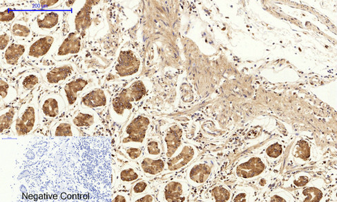
- Immunohistochemical analysis of paraffin-embedded Human-stomach tissue. 1,Calnexin Polyclonal Antibody was diluted at 1:200(4°C,overnight). 2, Sodium citrate pH 6.0 was used for antibody retrieval(>98°C,20min). 3,Secondary antibody was diluted at 1:200(room tempeRature, 30min). Negative control was used by secondary antibody only.
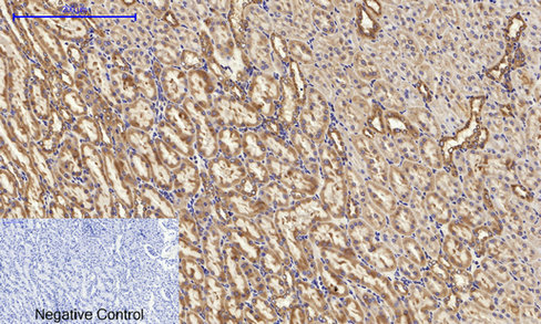
- Immunohistochemical analysis of paraffin-embedded Rat-kidney tissue. 1,Calnexin Polyclonal Antibody was diluted at 1:200(4°C,overnight). 2, Sodium citrate pH 6.0 was used for antibody retrieval(>98°C,20min). 3,Secondary antibody was diluted at 1:200(room tempeRature, 30min). Negative control was used by secondary antibody only.
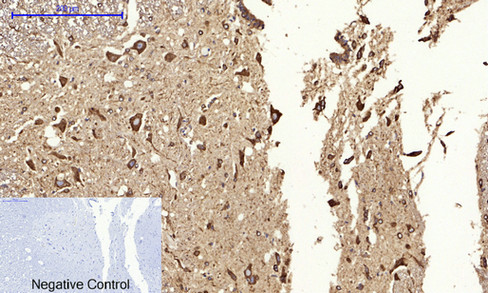
- Immunohistochemical analysis of paraffin-embedded Rat-spinal-cord tissue. 1,Calnexin Polyclonal Antibody was diluted at 1:200(4°C,overnight). 2, Sodium citrate pH 6.0 was used for antibody retrieval(>98°C,20min). 3,Secondary antibody was diluted at 1:200(room tempeRature, 30min). Negative control was used by secondary antibody only.
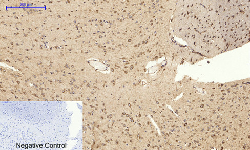
- Immunohistochemical analysis of paraffin-embedded Rat-brain tissue. 1,Calnexin Polyclonal Antibody was diluted at 1:200(4°C,overnight). 2, Sodium citrate pH 6.0 was used for antibody retrieval(>98°C,20min). 3,Secondary antibody was diluted at 1:200(room tempeRature, 30min). Negative control was used by secondary antibody only.
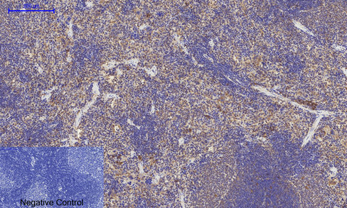
- Immunohistochemical analysis of paraffin-embedded Rat-spleen tissue. 1,Calnexin Polyclonal Antibody was diluted at 1:200(4°C,overnight). 2, Sodium citrate pH 6.0 was used for antibody retrieval(>98°C,20min). 3,Secondary antibody was diluted at 1:200(room tempeRature, 30min). Negative control was used by secondary antibody only.
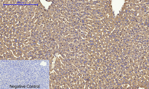
- Immunohistochemical analysis of paraffin-embedded Mouse-liver tissue. 1,Calnexin Polyclonal Antibody was diluted at 1:200(4°C,overnight). 2, Sodium citrate pH 6.0 was used for antibody retrieval(>98°C,20min). 3,Secondary antibody was diluted at 1:200(room tempeRature, 30min). Negative control was used by secondary antibody only.
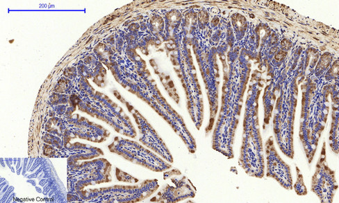
- Immunohistochemical analysis of paraffin-embedded Mouse-colon tissue. 1,Calnexin Polyclonal Antibody was diluted at 1:200(4°C,overnight). 2, Sodium citrate pH 6.0 was used for antibody retrieval(>98°C,20min). 3,Secondary antibody was diluted at 1:200(room tempeRature, 30min). Negative control was used by secondary antibody only.
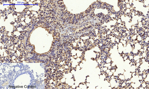
- Immunohistochemical analysis of paraffin-embedded Mouse-lung tissue. 1,Calnexin Polyclonal Antibody was diluted at 1:200(4°C,overnight). 2, Sodium citrate pH 6.0 was used for antibody retrieval(>98°C,20min). 3,Secondary antibody was diluted at 1:200(room tempeRature, 30min). Negative control was used by secondary antibody only.

- Immunohistochemical analysis of paraffin-embedded Mouse-kidney tissue. 1,Calnexin Polyclonal Antibody was diluted at 1:200(4°C,overnight). 2, Sodium citrate pH 6.0 was used for antibody retrieval(>98°C,20min). 3,Secondary antibody was diluted at 1:200(room tempeRature, 30min). Negative control was used by secondary antibody only.
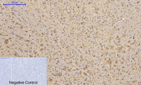
- Immunohistochemical analysis of paraffin-embedded Mouse-brain tissue. 1,Calnexin Polyclonal Antibody was diluted at 1:200(4°C,overnight). 2, Sodium citrate pH 6.0 was used for antibody retrieval(>98°C,20min). 3,Secondary antibody was diluted at 1:200(room tempeRature, 30min). Negative control was used by secondary antibody only.
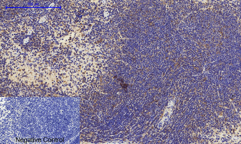
- Immunohistochemical analysis of paraffin-embedded Mouse-spleen tissue. 1,Calnexin Polyclonal Antibody was diluted at 1:200(4°C,overnight). 2, Sodium citrate pH 6.0 was used for antibody retrieval(>98°C,20min). 3,Secondary antibody was diluted at 1:200(room tempeRature, 30min). Negative control was used by secondary antibody only.
.jpg)
- Western Blot analysis of 293 cells using Calnexin Polyclonal Antibody diluted at 1:2000
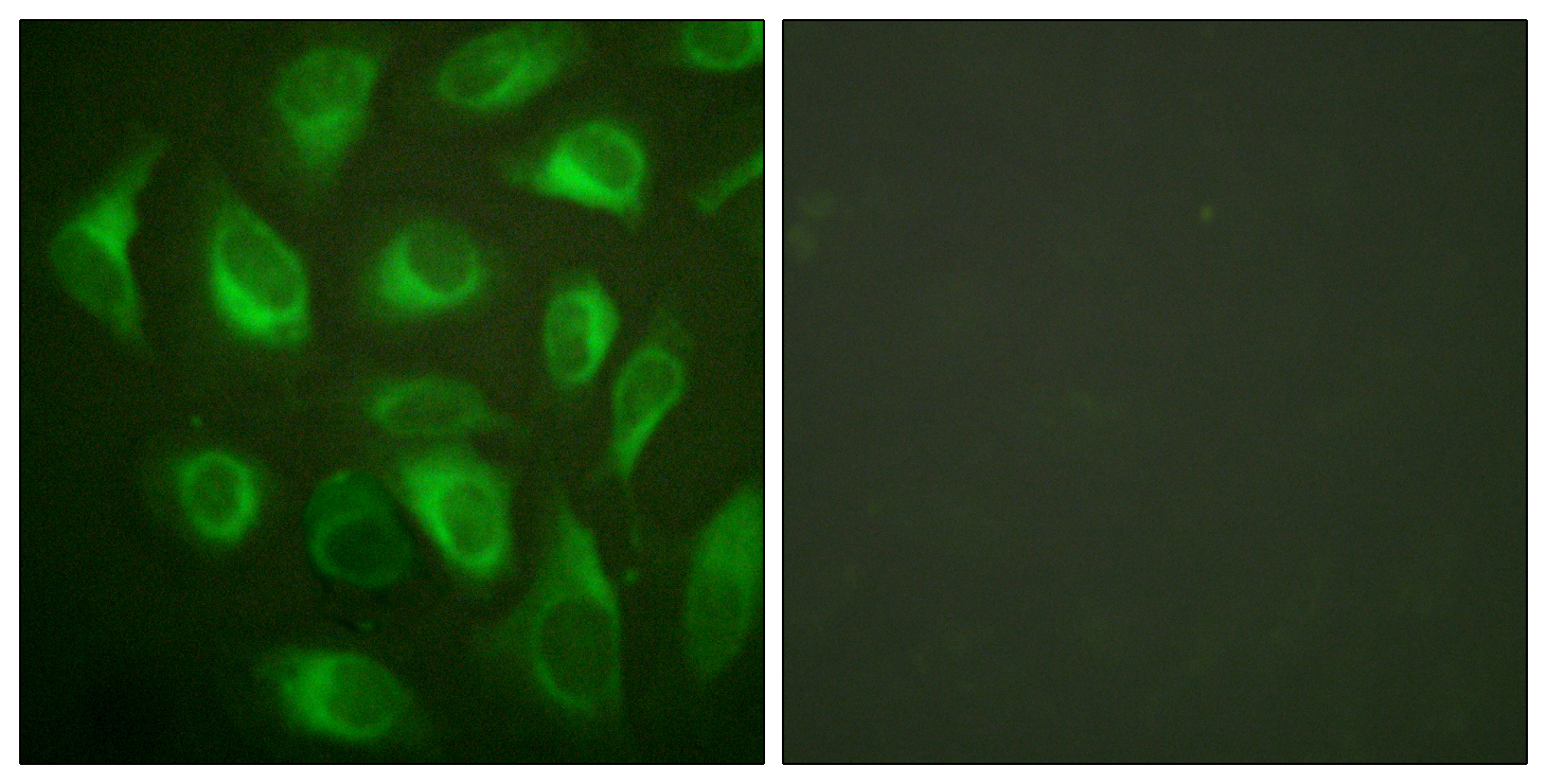
- Immunofluorescence analysis of HeLa cells, using Calnexin Antibody. The picture on the right is blocked with the synthesized peptide.

- Immunohistochemistry analysis of paraffin-embedded human lung carcinoma tissue, using Calnexin Antibody. The picture on the right is blocked with the synthesized peptide.
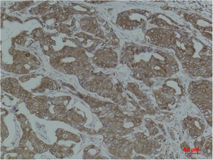
- Immunohistochemical analysis of paraffin-embedded Human Breast Carcinoma using Calnexin Polyclonal Antibody.
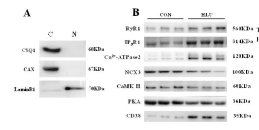
- New Findings: Hindlimb Unloading Causes Nucleocytoplasmic Ca2+ Overload and DNA Damage in Skeletal Muscle Cells Yunfang Gao WB Rat muscle cell


