Caspase12 Polyclonal Antibody
- Catalog No.:YT0654
- Applications:WB;IHC;IF;ELISA
- Reactivity:Human;Rat;Mouse;
- Target:
- Caspase-12
- Fields:
- >>Protein processing in endoplasmic reticulum;>>Apoptosis;>>NOD-like receptor signaling pathway;>>Alzheimer disease;>>Amyotrophic lateral sclerosis;>>Prion disease;>>Pathways of neurodegeneration - multiple diseases;>>Hepatitis B
- Gene Name:
- CASP12
- Protein Name:
- caspase12
- Human Gene Id:
- 120329
- Human Swiss Prot No:
- Q6UXS9
- Mouse Swiss Prot No:
- O08736
- Immunogen:
- The antiserum was produced against synthesized peptide derived from human Caspase12. AA range:50-99
- Specificity:
- Caspase12 Polyclonal Antibody detects endogenous levels of Caspase12 protein.
- Formulation:
- Liquid in PBS containing 50% glycerol, 0.5% BSA and 0.02% sodium azide.
- Source:
- Polyclonal, Rabbit,IgG
- Dilution:
- WB 1:500 - 1:2000. IHC 1:100 - 1:300. ELISA: 1:40000.. IF 1:50-200
- Purification:
- The antibody was affinity-purified from rabbit antiserum by affinity-chromatography using epitope-specific immunogen.
- Concentration:
- 1 mg/ml
- Storage Stability:
- -15°C to -25°C/1 year(Do not lower than -25°C)
- Other Name:
- CASP12;Inactive caspase-12;CASP-12
- Observed Band(KD):
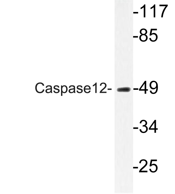
- 50kD
- Background:
- Caspases are cysteine proteases that cleave C-terminal aspartic acid residues on their substrate molecules. This gene is most highly related to members of the ICE subfamily of caspases that process inflammatory cytokines. In rodents, the homolog of this gene mediates apoptosis in response to endoplasmic reticulum stress. However, in humans this gene contains a polymorphism for the presence or absence of a premature stop codon. The majority of human individuals have the premature stop codon and produce a truncated non-functional protein. The read-through codon occurs primarily in individuals of African descent and carriers have endotoxin hypo-responsiveness and an increased susceptibility to severe sepsis. Several alternatively spliced transcript variants have been noted for this gene. [provided by RefSeq, Feb 2011],
- Function:
- proteolysis, apoptosis, virus-infected cell apoptosis, ER-nuclear signaling pathway, response to unfolded protein, cell death, response to organic substance, regulation of cell death, programmed cell death, death, endoplasmic reticulum unfolded protein response, cellular response to stress, cellular response to unfolded protein, response to endoplasmic reticulum stress, regulation of apoptosis, regulation of programmed cell death, response to protein stimulus,apoptosis in response to endoplasmic reticulum stress,
- Subcellular Location:
- endoplasmic reticulum,IPAF inflammasome complex,NLRP3 inflammasome complex,AIM2 inflammasome complex,
- Expression:
- Detected in heart, kidney, liver, lung, pancreas, small intestine, spleen, stomach, thymus and testis.
Antitumor Effect of Fluoxetine on Chronic Stress-Promoted Lung Cancer Growth via Suppressing Kynurenine Pathway and Enhancing Cellular Immunity. Frontiers in Pharmacology Front Pharmacol. 2021 Aug;0:1609 WB Mouse 1:2000 A549 cell-xenograft
Mitochondrial pathway and endoplasmic reticulum stress participate in the photosensitizing effectiveness of AE‐PDT in MG63 cells. Cancer Medicine 2016 Oct 03 WB Human MG63 cell
Endoplasmic reticulum stress plays an important role in methotrexate-related cognitive impairment in adult rats. International Journal of Clinical and Experimental Pathology Int J Clin Exp Patho. 2017; 10(10): 10252–10260 WB Rat 1:100 hippocampal tissues
Mitochondrial pathway and endoplasmic reticulum stress participate in the photosensitizing effectiveness of AE‐PDT in MG63 cells. Cancer Medicine 2016 Oct 03 WB Human MG63 cell
Augmented microglial endoplasmic reticulum-mitochondria contacts mediate depression-like behavior in mice induced by chronic social defeat stress Nature Communications Zhang Jia-Rui IHC Mouse 1:100 brain tissue
Dietary bitter ginger-derived zerumbone improved memory performance during aging through inhibition of the PERK/CHOP-dependent endoplasmic reticulum stress pathway Food & Function Chuan Yang WB Mouse,Human brain tissue SH-SY5Y cell
- June 19-2018
- WESTERN IMMUNOBLOTTING PROTOCOL
- June 19-2018
- IMMUNOHISTOCHEMISTRY-PARAFFIN PROTOCOL
- June 19-2018
- IMMUNOFLUORESCENCE PROTOCOL
- September 08-2020
- FLOW-CYTOMEYRT-PROTOCOL
- May 20-2022
- Cell-Based ELISA│解您多样本WB检测之困扰
- July 13-2018
- CELL-BASED-ELISA-PROTOCOL-FOR-ACETYL-PROTEIN
- July 13-2018
- CELL-BASED-ELISA-PROTOCOL-FOR-PHOSPHO-PROTEIN
- July 13-2018
- Antibody-FAQs
- Products Images

- Western Blot analysis of various cells using Caspase12 Polyclonal Antibody diluted at 1:500

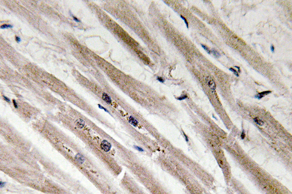
- Immunohistochemistry analysis of Caspase12 antibody in paraffin-embedded human heart tissue.

- Western blot analysis of lysate from HUVEC cells, using Caspase12 antibody.


