Cdc25A Polyclonal Antibody
- Catalog No.:YT0796
- Applications:WB;IHC;IF;ELISA
- Reactivity:Human;Mouse;Rat;Monkey
- Target:
- Cdc25A
- Fields:
- >>Cell cycle;>>Cellular senescence;>>Progesterone-mediated oocyte maturation;>>MicroRNAs in cancer;>>Chemical carcinogenesis - receptor activation
- Gene Name:
- CDC25A
- Protein Name:
- M-phase inducer phosphatase 1
- Human Gene Id:
- 993
- Human Swiss Prot No:
- P30304
- Mouse Swiss Prot No:
- P48964
- Rat Gene Id:
- 171102
- Rat Swiss Prot No:
- P48965
- Immunogen:
- The antiserum was produced against synthesized peptide derived from human CDC25A. AA range:43-92
- Specificity:
- Cdc25A Polyclonal Antibody detects endogenous levels of Cdc25A protein.
- Formulation:
- Liquid in PBS containing 50% glycerol, 0.5% BSA and 0.02% sodium azide.
- Source:
- Polyclonal, Rabbit,IgG
- Dilution:
- WB 1:500 - 1:2000. IHC 1:100 - 1:300. IF 1:200 - 1:1000. ELISA: 1:10000. Not yet tested in other applications.
- Purification:
- The antibody was affinity-purified from rabbit antiserum by affinity-chromatography using epitope-specific immunogen.
- Concentration:
- 1 mg/ml
- Storage Stability:
- -15°C to -25°C/1 year(Do not lower than -25°C)
- Other Name:
- CDC25A;M-phase inducer phosphatase 1;Dual specificity phosphatase Cdc25A
- Observed Band(KD):
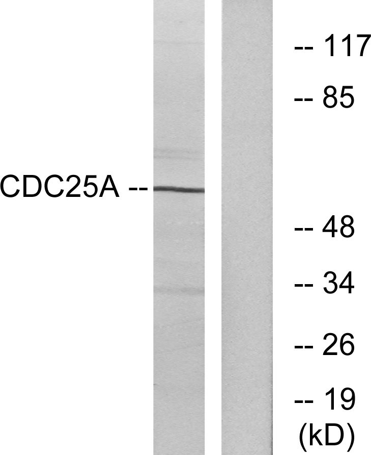
- 60kD
- Background:
- cell division cycle 25A(CDC25A) Homo sapiens CDC25A is a member of the CDC25 family of phosphatases. CDC25A is required for progression from G1 to the S phase of the cell cycle. It activates the cyclin-dependent kinase CDC2 by removing two phosphate groups. CDC25A is specifically degraded in response to DNA damage, which prevents cells with chromosomal abnormalities from progressing through cell division. CDC25A is an oncogene, although its exact role in oncogenesis has not been demonstrated. Two transcript variants encoding different isoforms have been found for this gene. [provided by RefSeq, Jul 2008],
- Function:
- catalytic activity:Protein tyrosine phosphate + H(2)O = protein tyrosine + phosphate.,domain:The phosphodegron motif mediates interaction with specific F-box proteins when phosphorylated. Putative phosphorylation sites at Ser-79 and Ser-82 appear to be essential for this interaction.,enzyme regulation:Stimulated by B-type cyclins.,function:Tyrosine protein phosphatase which functions as a dosage-dependent inducer of mitotic progression. Directly dephosphorylates CDC2 and stimulates its kinase activity. Also dephosphorylates CDK2 in complex with cyclin E, in vitro.,PTM:Phosphorylated by CHEK1 on Ser-76, Ser-124, Ser-178, Ser-279, Ser-293 and Thr-507 during checkpoint mediated cell cycle arrest. Also phosphorylated by CHEK2 on Ser-124, Ser-279, and Ser-293 during checkpoint mediated cell cycle arrest. Phosphorylation on Ser-178 and Thr-507 creates binding sites for YWHAE/14-3-3 epsilon whi
- Subcellular Location:
- intracellular,nucleus,nucleoplasm,cytoplasm,cytosol,
- Expression:
- Lymph,
- June 19-2018
- WESTERN IMMUNOBLOTTING PROTOCOL
- June 19-2018
- IMMUNOHISTOCHEMISTRY-PARAFFIN PROTOCOL
- June 19-2018
- IMMUNOFLUORESCENCE PROTOCOL
- September 08-2020
- FLOW-CYTOMEYRT-PROTOCOL
- May 20-2022
- Cell-Based ELISA│解您多样本WB检测之困扰
- July 13-2018
- CELL-BASED-ELISA-PROTOCOL-FOR-ACETYL-PROTEIN
- July 13-2018
- CELL-BASED-ELISA-PROTOCOL-FOR-PHOSPHO-PROTEIN
- July 13-2018
- Antibody-FAQs
- Products Images

- Li, Lin, et al. "Telekin suppresses human hepatocellular carcinoma cells in vitro by inducing G 2/M phase arrest via the p38 MAPK signaling pathway." Acta Pharmacologica Sinica 35.10 (2014): 1311.

- Western blot analysis of lysates from A2780 cells, treated with UV, using CDC25A Antibody. The lane on the right is blocked with the synthesized peptide.

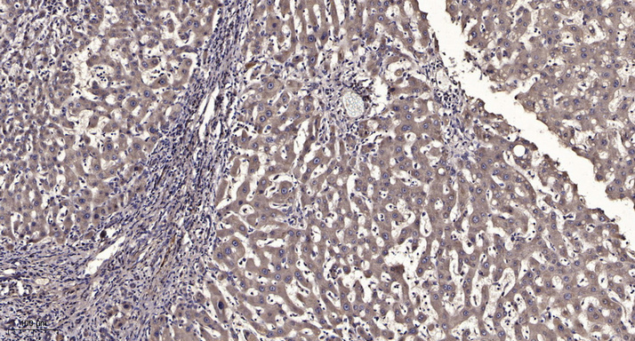
- Immunohistochemical analysis of paraffin-embedded human liver cancer. 1, Antibody was diluted at 1:200(4° overnight). 2, Tris-EDTA,pH9.0 was used for antigen retrieval. 3,Secondary antibody was diluted at 1:200(room temperature, 45min).


