CIDE-3 Polyclonal Antibody
- Catalog No.:YT0927
- Applications:IHC;IF;ELISA
- Reactivity:Human;Rat;Mouse;
- Target:
- CIDE-3
- Gene Name:
- CIDEC
- Protein Name:
- Cell death activator CIDE-3
- Human Gene Id:
- 63924
- Human Swiss Prot No:
- Q96AQ7
- Mouse Swiss Prot No:
- P56198
- Immunogen:
- The antiserum was produced against synthesized peptide derived from human CIDEC. AA range:189-238
- Specificity:
- CIDE-3 Polyclonal Antibody detects endogenous levels of CIDE-3 protein.
- Formulation:
- Liquid in PBS containing 50% glycerol, 0.5% BSA and 0.02% sodium azide.
- Source:
- Polyclonal, Rabbit,IgG
- Dilution:
- IHC 1:100 - 1:300. IF 1:200 - 1:1000. ELISA: 1:5000. Not yet tested in other applications.
- Purification:
- The antibody was affinity-purified from rabbit antiserum by affinity-chromatography using epitope-specific immunogen.
- Concentration:
- 1 mg/ml
- Storage Stability:
- -15°C to -25°C/1 year(Do not lower than -25°C)
- Other Name:
- CIDEC;FSP27;Cell death activator CIDE-3;Cell death-inducing DFFA-like effector protein C;Fat-specific protein FSP27 homolog
- Molecular Weight(Da):
- 27kD
- Background:
- cell death inducing DFFA like effector c(CIDEC) Homo sapiens This gene encodes a member of the cell death-inducing DNA fragmentation factor-like effector family. Members of this family play important roles in apoptosis. The encoded protein promotes lipid droplet formation in adipocytes and may mediate adipocyte apoptosis. This gene is regulated by insulin and its expression is positively correlated with insulin sensitivity. Mutations in this gene may contribute to insulin resistant diabetes. A pseudogene of this gene is located on the short arm of chromosome 3. Alternatively spliced transcript variants that encode different isoforms have been observed for this gene. [provided by RefSeq, Dec 2010],
- Function:
- function:Isoforms 1 and 2 induce apoptosis.,similarity:Contains 1 CIDE-N domain.,subcellular location:Cytoplasmic in a punctate manner.,tissue specificity:Expressed mainly in small intestine, heart, colon and stomach and, at lower levels, in brain, kidney and liver.,
- Subcellular Location:
- Nucleus . Endoplasmic reticulum. Lipid droplet. Diffuses quickly on lipid droplet surface, but becomes trapped and clustered at lipid droplet contact sites, thereby enabling its rapid enrichment at lipid droplet contact sites.
- Expression:
- Expressed mainly in adipose tissue, small intestine, heart, colon and stomach and, at lower levels, in brain, kidney and liver.
- June 19-2018
- WESTERN IMMUNOBLOTTING PROTOCOL
- June 19-2018
- IMMUNOHISTOCHEMISTRY-PARAFFIN PROTOCOL
- June 19-2018
- IMMUNOFLUORESCENCE PROTOCOL
- September 08-2020
- FLOW-CYTOMEYRT-PROTOCOL
- May 20-2022
- Cell-Based ELISA│解您多样本WB检测之困扰
- July 13-2018
- CELL-BASED-ELISA-PROTOCOL-FOR-ACETYL-PROTEIN
- July 13-2018
- CELL-BASED-ELISA-PROTOCOL-FOR-PHOSPHO-PROTEIN
- July 13-2018
- Antibody-FAQs
- Products Images

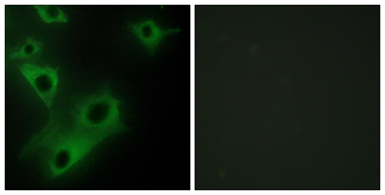
- Immunofluorescence analysis of HeLa cells, using CIDEC Antibody. The picture on the right is blocked with the synthesized peptide.

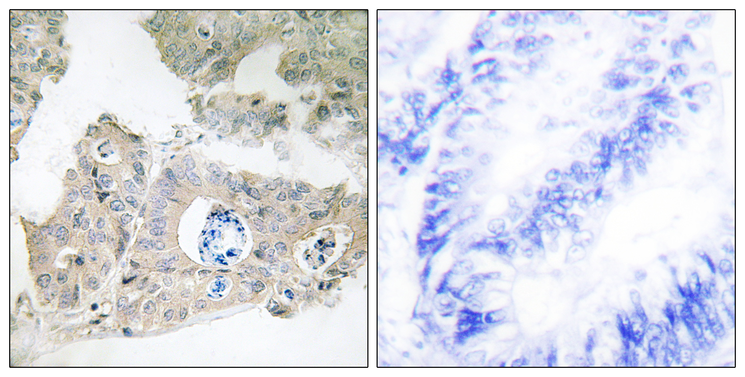
- Immunohistochemistry analysis of paraffin-embedded human colon carcinoma tissue, using CIDEC Antibody. The picture on the right is blocked with the synthesized peptide.


