COL5A1 Polyclonal Antibody
- Catalog No.:YT1030
- Applications:WB;IHC;IF;ELISA
- Reactivity:Human;Rat;Mouse;
- Target:
- COL5A1
- Fields:
- >>Protein digestion and absorption
- Gene Name:
- COL5A1
- Protein Name:
- Collagen alpha-1(V) chain
- Human Gene Id:
- 1289
- Human Swiss Prot No:
- P20908
- Mouse Swiss Prot No:
- O88207
- Immunogen:
- The antiserum was produced against synthesized peptide derived from human Collagen V alpha1. AA range:301-350
- Specificity:
- COL5A1 Polyclonal Antibody detects endogenous levels of COL5A1 protein.
- Formulation:
- Liquid in PBS containing 50% glycerol, 0.5% BSA and 0.02% sodium azide.
- Source:
- Polyclonal, Rabbit,IgG
- Dilution:
- WB 1:500 - 1:2000. IHC 1:100 - 1:300. IF 1:200 - 1:1000. ELISA: 1:20000. Not yet tested in other applications.
- Purification:
- The antibody was affinity-purified from rabbit antiserum by affinity-chromatography using epitope-specific immunogen.
- Concentration:
- 1 mg/ml
- Storage Stability:
- -15°C to -25°C/1 year(Do not lower than -25°C)
- Other Name:
- COL5A1;Collagen alpha-1(V) chain
- Observed Band(KD):
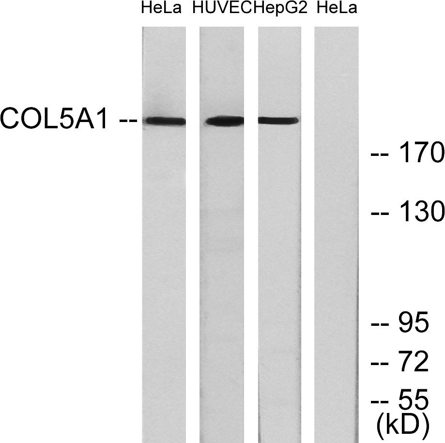
- 200kD
- Background:
- This gene encodes an alpha chain for one of the low abundance fibrillar collagens. Fibrillar collagen molecules are trimers that can be composed of one or more types of alpha chains. Type V collagen is found in tissues containing type I collagen and appears to regulate the assembly of heterotypic fibers composed of both type I and type V collagen. This gene product is closely related to type XI collagen and it is possible that the collagen chains of types V and XI constitute a single collagen type with tissue-specific chain combinations. The encoded procollagen protein occurs commonly as the heterotrimer pro-alpha1(V)-pro-alpha1(V)-pro-alpha2(V). Mutations in this gene are associated with Ehlers-Danlos syndrome, types I and II. Alternative splicing of this gene results in multiple transcript variants. [provided by RefSeq, May 2013],
- Function:
- disease:Defects in COL5A1 are a cause of Ehlers-Danlos syndrome type 1 (EDS1) [MIM:130000]; also known as Ehlers-Danlos syndrome gravis or severe classic type Ehlers-Danlos syndrome. EDS is a connective tissue disorder characterized by hyperextensible skin, atrophic cutaneous scars due to tissue fragility and joint hyperlaxity. EDS1 is the severe form of classic Ehlers-Danlos syndrome.,disease:Defects in COL5A1 are a cause of Ehlers-Danlos syndrome type 2 (EDS2) [MIM:130010]; also known as Ehlers-Danlos syndrome mitis or mild classic type Ehlers Danlos syndrome.,function:Type V collagen is a member of group I collagen (fibrillar forming collagen). It is a minor connective tissue component of nearly ubiquitous distribution. Type V collagen binds to DNA, heparan sulfate, thrombospondin, heparin, and insulin.,PTM:Prolines at the third position of the tripeptide repeating unit (G-X-Y) are hy
- Subcellular Location:
- Secreted, extracellular space, extracellular matrix .
- Expression:
- Aorta endothelial cell,Chorioamniotic membrane,Eye,Placenta,
Myofibroblasts derived type V collagen promoting tissue mechanical stress and facilitating metastasis and therapy resistance of lung adenocarcinoma cells Cell Death & Disease Zhu Guangsheng IHC,IF Human 1:200 Lung Adenocarcinoma (LUAD) tissue cancer-associated fibroblasts (CAFs)
- June 19-2018
- WESTERN IMMUNOBLOTTING PROTOCOL
- June 19-2018
- IMMUNOHISTOCHEMISTRY-PARAFFIN PROTOCOL
- June 19-2018
- IMMUNOFLUORESCENCE PROTOCOL
- September 08-2020
- FLOW-CYTOMEYRT-PROTOCOL
- May 20-2022
- Cell-Based ELISA│解您多样本WB检测之困扰
- July 13-2018
- CELL-BASED-ELISA-PROTOCOL-FOR-ACETYL-PROTEIN
- July 13-2018
- CELL-BASED-ELISA-PROTOCOL-FOR-PHOSPHO-PROTEIN
- July 13-2018
- Antibody-FAQs
- Products Images

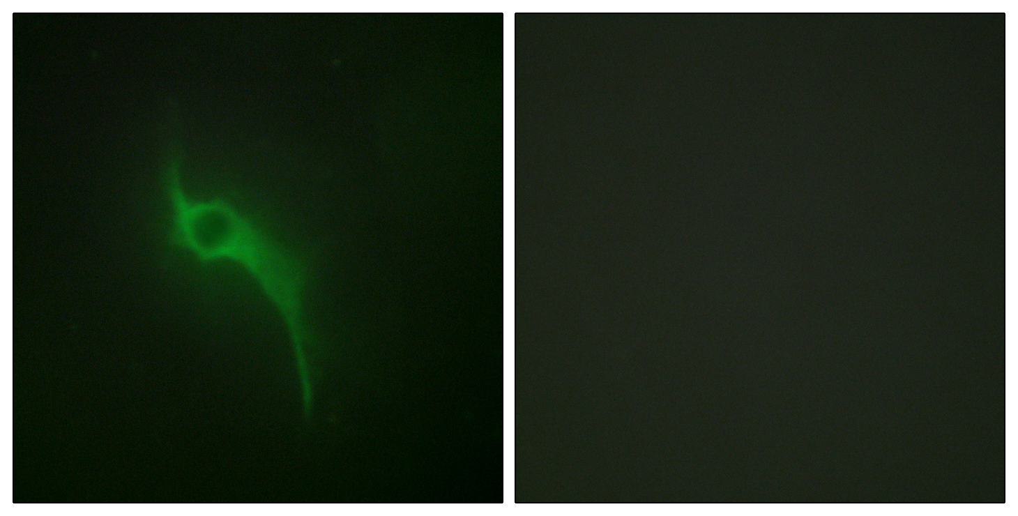
- Immunofluorescence analysis of HeLa cells, using Collagen V alpha1 Antibody. The picture on the right is blocked with the synthesized peptide.

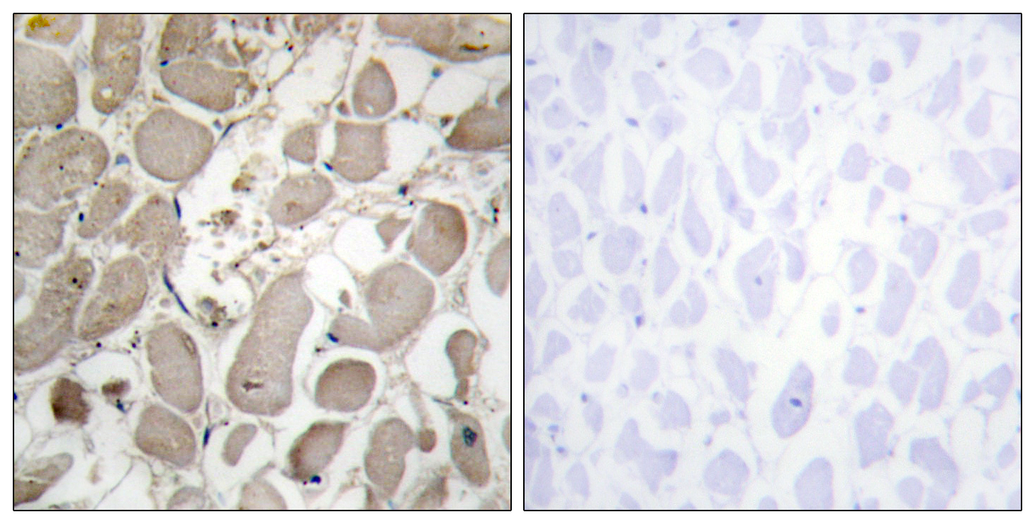
- Immunohistochemistry analysis of paraffin-embedded human heart tissue, using Collagen V alpha1 Antibody. The picture on the right is blocked with the synthesized peptide.

- Western blot analysis of lysates from HeLa, and HUVEC, and HepG2 cells, using Collagen V alpha1 Antibody. The lane on the right is blocked with the synthesized peptide.


