DAP-5 Polyclonal Antibody
- Catalog No.:YT1286
- Applications:WB;IHC;IF;ELISA
- Reactivity:Human;Mouse
- Target:
- DAP-5
- Fields:
- >>Viral myocarditis
- Gene Name:
- EIF4G2
- Protein Name:
- Eukaryotic translation initiation factor 4 gamma 2
- Human Gene Id:
- 1982
- Human Swiss Prot No:
- P78344
- Mouse Gene Id:
- 13690
- Mouse Swiss Prot No:
- Q62448
- Immunogen:
- The antiserum was produced against synthesized peptide derived from human EIF4G2. AA range:41-90
- Specificity:
- DAP-5 Polyclonal Antibody detects endogenous levels of DAP-5 protein.
- Formulation:
- Liquid in PBS containing 50% glycerol, 0.5% BSA and 0.02% sodium azide.
- Source:
- Polyclonal, Rabbit,IgG
- Dilution:
- WB 1:500 - 1:2000. IHC 1:100 - 1:300. IF 1:200 - 1:1000. ELISA: 1:20000. Not yet tested in other applications.
- Purification:
- The antibody was affinity-purified from rabbit antiserum by affinity-chromatography using epitope-specific immunogen.
- Concentration:
- 1 mg/ml
- Storage Stability:
- -15°C to -25°C/1 year(Do not lower than -25°C)
- Other Name:
- EIF4G2;DAP5;OK/SW-cl.75;Eukaryotic translation initiation factor 4 gamma 2;eIF-4-gamma 2;eIF-4G 2;eIF4G 2;Death-associated protein 5;DAP-5;p97
- Observed Band(KD):
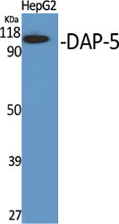
- 90kD
- Background:
- Translation initiation is mediated by specific recognition of the cap structure by eukaryotic translation initiation factor 4F (eIF4F), which is a cap binding protein complex that consists of three subunits: eIF4A, eIF4E and eIF4G. The protein encoded by this gene shares similarity with the C-terminal region of eIF4G that contains the binding sites for eIF4A and eIF3; eIF4G, in addition, contains a binding site for eIF4E at the N-terminus. Unlike eIF4G, which supports cap-dependent and independent translation, this gene product functions as a general repressor of translation by forming translationally inactive complexes. In vitro and in vivo studies indicate that translation of this mRNA initiates exclusively at a non-AUG (GUG) codon. Alternatively spliced transcript variants encoding different isoforms of this gene have been described. [provided by RefSeq, Jul 2008],
- Function:
- function:Appears to play a role in the switch from cap-dependent to IRES-mediated translation during mitosis, apoptosis and viral infection. Cleaved by some caspases and viral proteases.,miscellaneous:This gene has been shown to be extensively edited in the liver of APOBEC1 transgenic animal model. Its aberrant editing could contribute to the potent oncogenesis induced by overexpression of APOBEC1. The aberrant edited sequence, called NAT1, is likely to be a fundamental translational repressor.,PTM:Phosphorylation; hyperphosphorylated during mitosis.,similarity:Belongs to the eIF4G family.,similarity:Contains 1 MI domain.,similarity:Contains 1 MIF4G domain.,similarity:Contains 1 W2 domain.,subunit:Interacts with the serine/threonine protein kinases MKNK1 and MKNK2. Binds EIF4A and EIF3. Interacts with MIF4GD.,tissue specificity:Ubiquitously expressed in all adult tissues examined, with h
- Subcellular Location:
- cytosol,cell-cell adherens junction,membrane,eukaryotic translation initiation factor 4F complex,axon,
- Expression:
- Ubiquitously expressed in all adult tissues examined, with high levels in skeletal muscle and heart. Also expressed in fetal brain, lung, liver and kidney.
- June 19-2018
- WESTERN IMMUNOBLOTTING PROTOCOL
- June 19-2018
- IMMUNOHISTOCHEMISTRY-PARAFFIN PROTOCOL
- June 19-2018
- IMMUNOFLUORESCENCE PROTOCOL
- September 08-2020
- FLOW-CYTOMEYRT-PROTOCOL
- May 20-2022
- Cell-Based ELISA│解您多样本WB检测之困扰
- July 13-2018
- CELL-BASED-ELISA-PROTOCOL-FOR-ACETYL-PROTEIN
- July 13-2018
- CELL-BASED-ELISA-PROTOCOL-FOR-PHOSPHO-PROTEIN
- July 13-2018
- Antibody-FAQs
- Products Images

- Western Blot analysis of various cells using DAP-5 Polyclonal Antibody
.jpg)
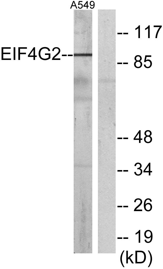
- Western Blot analysis of A549 cells using DAP-5 Polyclonal Antibody

- Western blot analysis of lysates from A549 cells, using EIF4G2 Antibody. The lane on the right is blocked with the synthesized peptide.

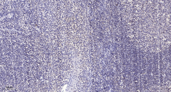
- Immunohistochemical analysis of paraffin-embedded human tonsil. 1, Antibody was diluted at 1:200(4° overnight). 2, Tris-EDTA,pH9.0 was used for antigen retrieval. 3,Secondary antibody was diluted at 1:200(room temperature, 30min).


