Daxx Polyclonal Antibody
- Catalog No.:YT1292
- Applications:WB;IHC;IF;ELISA
- Reactivity:Human;Mouse;Rat
- Target:
- Daxx
- Fields:
- >>MAPK signaling pathway;>>Apoptosis;>>Parkinson disease;>>Amyotrophic lateral sclerosis;>>Pathways of neurodegeneration - multiple diseases;>>Herpes simplex virus 1 infection
- Gene Name:
- DAXX
- Protein Name:
- Death domain-associated protein 6
- Human Gene Id:
- 1616
- Human Swiss Prot No:
- Q9UER7
- Mouse Gene Id:
- 13163
- Mouse Swiss Prot No:
- O35613
- Rat Swiss Prot No:
- Q8VIB2
- Immunogen:
- The antiserum was produced against synthesized peptide derived from human DAXX. AA range:361-410
- Specificity:
- Daxx Polyclonal Antibody detects endogenous levels of Daxx protein.
- Formulation:
- Liquid in PBS containing 50% glycerol, 0.5% BSA and 0.02% sodium azide.
- Source:
- Polyclonal, Rabbit,IgG
- Dilution:
- WB 1:500 - 1:2000. IHC 1:100 - 1:300. IF 1:200 - 1:1000. ELISA: 1:20000. Not yet tested in other applications.
- Purification:
- The antibody was affinity-purified from rabbit antiserum by affinity-chromatography using epitope-specific immunogen.
- Concentration:
- 1 mg/ml
- Storage Stability:
- -15°C to -25°C/1 year(Do not lower than -25°C)
- Other Name:
- DAXX;BING2;DAP6;Death domain-associated protein 6;Daxx;hDaxx;ETS1-associated protein 1;EAP1;Fas death domain-associated protein
- Observed Band(KD):
- 85-115kd
- Background:
- This gene encodes a multifunctional protein that resides in multiple locations in the nucleus and in the cytoplasm. It interacts with a wide variety of proteins, such as apoptosis antigen Fas, centromere protein C, and transcription factor erythroblastosis virus E26 oncogene homolog 1. In the nucleus, the encoded protein functions as a potent transcription repressor that binds to sumoylated transcription factors. Its repression can be relieved by the sequestration of this protein into promyelocytic leukemia nuclear bodies or nucleoli. This protein also associates with centromeres in G2 phase. In the cytoplasm, the encoded protein may function to regulate apoptosis. The subcellular localization and function of this protein are modulated by post-translational modifications, including sumoylation, phosphorylation and polyubiquitination. Alternative splicing results in multiple transcript varian
- Function:
- function:Proposed to mediate activation of the JNK pathway and apoptosis via MAP3K5 in response to signaling from TNFRSF6 and TGFBR2. Interaction with HSPB1/HSP27 may prevent interaction with TNFRSF6 and MAP3K5 and block DAXX-mediated apoptosis. In contrast, in lymphoid cells JNC activation and TNFRSF6-mediated apoptosis may not involve DAXX. Seems to regulate transcription in PML/POD/ND10 nuclear bodies together with PML and may influence TNFRSF6-dependent apoptosis thereby. Down-regulates basal and activated transcription. Seems to act as a transcriptional co-repressor and inhibits PAX3 and ETS1 through direct protein-protein interaction. Modulates PAX5 activity. Its transcription repressor activity is modulated by recruiting it to subnuclear compartments like the nucleolus or PML/POD/ND10 nuclear bodies through interactions with MCSR1 and PML, respectively.,induction:Upon mitogenic st
- Subcellular Location:
- Cytoplasm . Nucleus, nucleoplasm . Nucleus, PML body . Nucleus, nucleolus . Chromosome, centromere . Dispersed throughout the nucleoplasm, in PML/POD/ND10 nuclear bodies, and in nucleoli (Probable). Colocalizes with histone H3.3, ATRX, HIRA and ASF1A at PML-nuclear bodies (PubMed:12953102, PubMed:14990586, PubMed:23222847, PubMed:24200965). Colocalizes with a subset of interphase centromeres, but is absent from mitotic centromeres (PubMed:9645950). Detected in cytoplasmic punctate structures (PubMed:11842083). Translocates from the nucleus to the cytoplasm upon glucose deprivation or oxidative stress (PubMed:12968034). Colocalizes with RASSF1 in the nucleus (PubMed:18566590). Colocalizes with USP7 in nucleoplasma with accumulation in speckled structures (PubMed:16845383). .; [Isoform beta]
- Expression:
- Ubiquitous.
- June 19-2018
- WESTERN IMMUNOBLOTTING PROTOCOL
- June 19-2018
- IMMUNOHISTOCHEMISTRY-PARAFFIN PROTOCOL
- June 19-2018
- IMMUNOFLUORESCENCE PROTOCOL
- September 08-2020
- FLOW-CYTOMEYRT-PROTOCOL
- May 20-2022
- Cell-Based ELISA│解您多样本WB检测之困扰
- July 13-2018
- CELL-BASED-ELISA-PROTOCOL-FOR-ACETYL-PROTEIN
- July 13-2018
- CELL-BASED-ELISA-PROTOCOL-FOR-PHOSPHO-PROTEIN
- July 13-2018
- Antibody-FAQs
- Products Images
.jpg)
- Western Blot analysis of A549 cells using Daxx Polyclonal Antibody

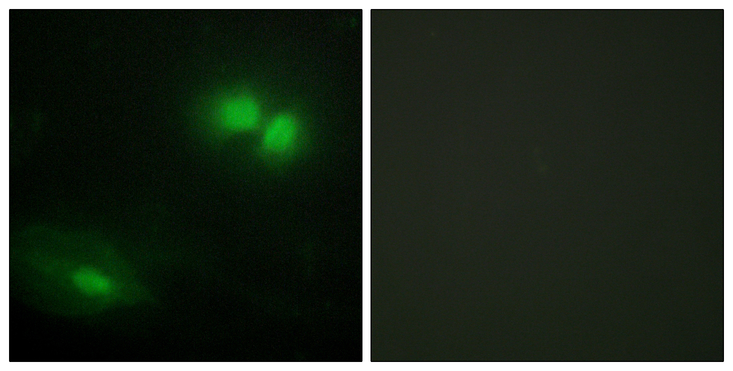
- Immunofluorescence analysis of HeLa cells, using DAXX Antibody. The picture on the right is blocked with the synthesized peptide.

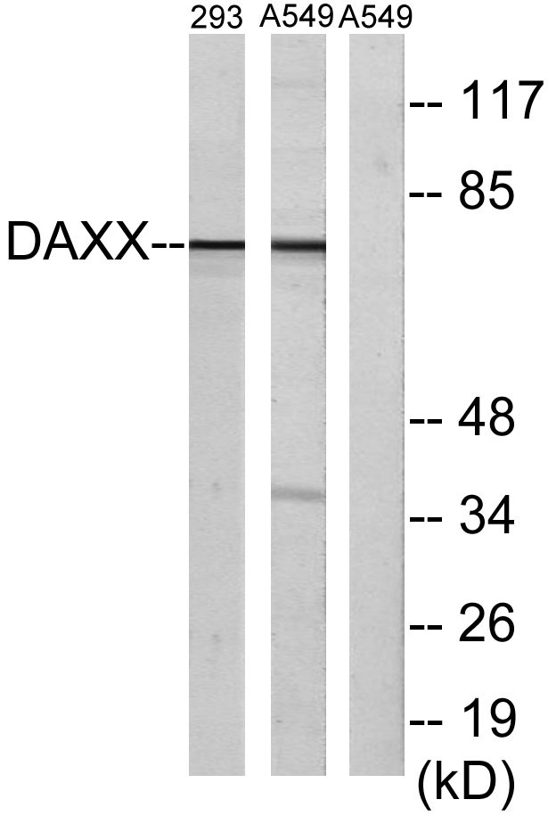
- Western blot analysis of lysates from 293 cells and A549 cells, using DAXX Antibody. The lane on the right is blocked with the synthesized peptide.

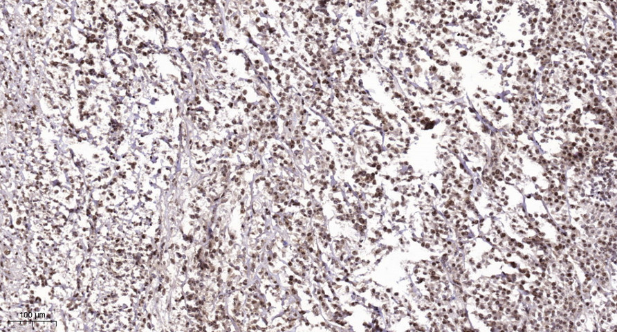
- Immunohistochemical analysis of paraffin-embedded human Colon cancer. 1, Antibody was diluted at 1:200(4° overnight). 2, Tris-EDTA,pH9.0 was used for antigen retrieval. 3,Secondary antibody was diluted at 1:200(room temperature, 45min).


