Dio-1 Polyclonal Antibody
- Catalog No.:YT1351
- Applications:WB;IHC;IF;ELISA
- Reactivity:Human;Mouse
- Target:
- Dio-1
- Gene Name:
- DIDO1
- Protein Name:
- Death-inducer obliterator 1
- Human Gene Id:
- 11083
- Human Swiss Prot No:
- Q9BTC0
- Mouse Gene Id:
- 23856
- Mouse Swiss Prot No:
- Q8C9B9
- Immunogen:
- The antiserum was produced against synthesized peptide derived from human DIDO1. AA range:161-210
- Specificity:
- Dio-1 Polyclonal Antibody detects endogenous levels of Dio-1 protein.
- Formulation:
- Liquid in PBS containing 50% glycerol, 0.5% BSA and 0.02% sodium azide.
- Source:
- Polyclonal, Rabbit,IgG
- Dilution:
- WB 1:500 - 1:2000. IHC 1:100 - 1:300. ELISA: 1:10000.. IF 1:50-200
- Purification:
- The antibody was affinity-purified from rabbit antiserum by affinity-chromatography using epitope-specific immunogen.
- Concentration:
- 1 mg/ml
- Storage Stability:
- -15°C to -25°C/1 year(Do not lower than -25°C)
- Other Name:
- DIDO1;C20orf158;DATF1;KIAA0333;Death-inducer obliterator 1;DIO-1;hDido1;Death-associated transcription factor 1;DATF-1
- Observed Band(KD):
- 244kD
- Background:
- Apoptosis, a major form of cell death, is an efficient mechanism for eliminating unwanted cells and is of central importance for development and homeostasis in metazoan animals. In mice, the death inducer-obliterator-1 gene is upregulated by apoptotic signals and encodes a cytoplasmic protein that translocates to the nucleus upon apoptotic signal activation. When overexpressed, the mouse protein induced apoptosis in cell lines growing in vitro. This gene is similar to the mouse gene and therefore is thought to be involved in apoptosis. Alternatively spliced transcripts have been found for this gene, encoding multiple isoforms. [provided by RefSeq, Jul 2008],
- Function:
- disease:Defects in DIDO1 may be a cause of myeloid neoplasms.,function:Putative transcription factor, weakly pro-apoptotic when overexpressed (By similarity). Tumor suppressor.,similarity:Contains 1 PHD-type zinc finger.,similarity:Contains 1 TFIIS central domain.,subcellular location:Translocates to the nucleus after pro-apoptotic stimuli.,tissue specificity:Ubiquitous.,
- Subcellular Location:
- Cytoplasm . Nucleus . Cytoplasm, cytoskeleton, spindle . Translocates to the nucleus after pro-apoptotic stimuli (By similarity). Translocates to the mitotic spindle upon loss of interaction with H3K4me3 during early mitosis. .
- Expression:
- Ubiquitous.
Chronic restraint stress induced changes in colonic homeostasis-related indexes and tryptophan-kynurenine metabolism in rats. Journal of Proteomics J Proteomics. 2021 May;240:104190 WB Rat 1 : 1000 Colon
- June 19-2018
- WESTERN IMMUNOBLOTTING PROTOCOL
- June 19-2018
- IMMUNOHISTOCHEMISTRY-PARAFFIN PROTOCOL
- June 19-2018
- IMMUNOFLUORESCENCE PROTOCOL
- September 08-2020
- FLOW-CYTOMEYRT-PROTOCOL
- May 20-2022
- Cell-Based ELISA│解您多样本WB检测之困扰
- July 13-2018
- CELL-BASED-ELISA-PROTOCOL-FOR-ACETYL-PROTEIN
- July 13-2018
- CELL-BASED-ELISA-PROTOCOL-FOR-PHOSPHO-PROTEIN
- July 13-2018
- Antibody-FAQs
- Products Images

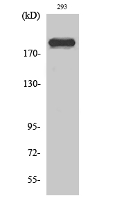
- Western Blot analysis of various cells using Dio-1 Polyclonal Antibody diluted at 1:2000

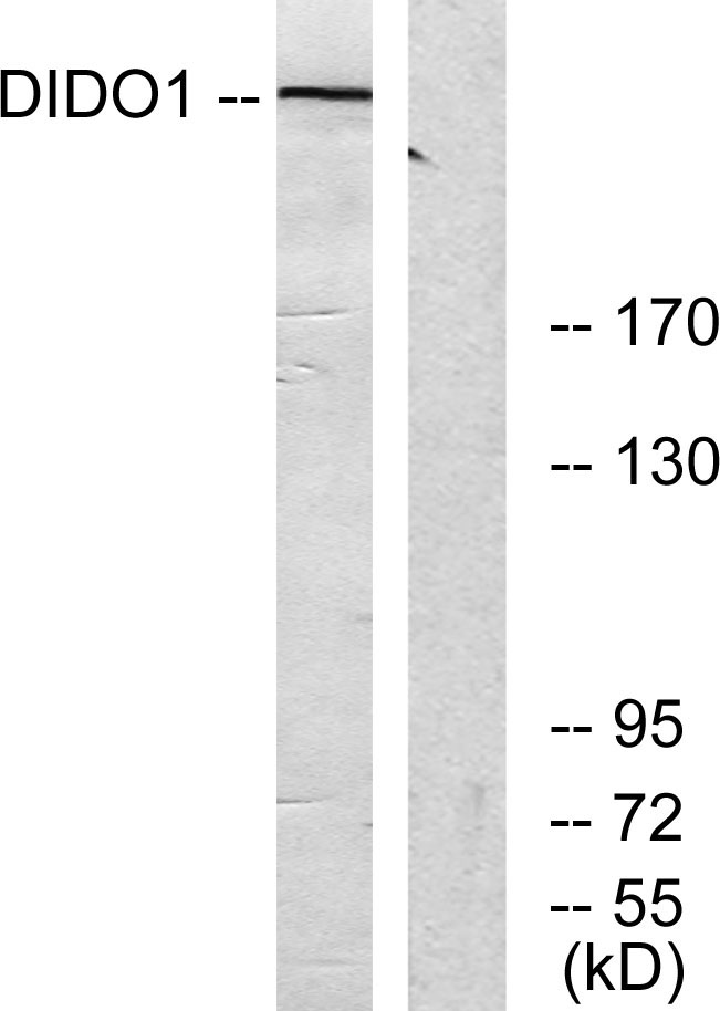
- Western blot analysis of lysates from 293 cells, using DIDO1 Antibody. The lane on the right is blocked with the synthesized peptide.

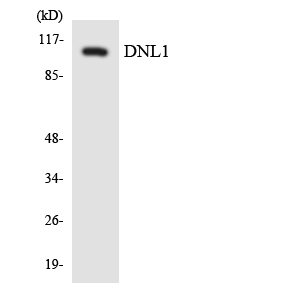
- Western blot analysis of the lysates from HepG2 cells using DNL1 antibody.

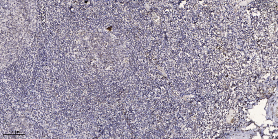
- Immunohistochemical analysis of paraffin-embedded human tonsil. 1, Antibody was diluted at 1:200(4° overnight). 2, Tris-EDTA,pH9.0 was used for antigen retrieval. 3,Secondary antibody was diluted at 1:200(room temperature, 45min).


